Category

Surgery:

Category

Wounds can be extremely painful. These wounds can also pose a health risk if they become inflamed or infected. The cause of the suture getting infected could be bacteria, especially those that are naturally found on the skin and Atmosphere. Thus, wound closure may end up promoting bacteria proliferation which can lead to wound complications. As a result, operating doctors, nurses, and other medical professionals have created ways to reduce surgical site infection, speed up healing, and prevent any of these healthcare-associated infections (HCAIs) and surgical site infections (SSIs) from spreading further. One of the ways they have done this is by using antibacterial sutures (ABS) in wound. This closure procedures article explores the advantages of using antibacterial sutures in wound closure procedures. There are various ways and techniques to treat wounds – sutures, staples, non-surgical medications, etc. 

What Is a Suture?

A suture, also known as a stitch, is a very thin, long thread that is twisted together to form a single strand in case of braided structure or comprises of single thread called Monofilament. Suturing is a method of tying off blood vessels to prevent bleeding and hold a wound together till the natural healing process is sufficiently established.

What is an Antibacterial Suture?

Any wound, by and large, are may get contaminated at the time of closure. As a solution to this problem, an antibacterial suture is a surgical innovation which reduces the risk of Surgical Site Infection (SSI). Antibacterial sutures are coated with Antibacterial agents and other agents containing antibacterial properties. They may help in healing wounds faster by significantly reducing the risk of surgical site infections. Why do you need to use an antibacterial suture? We need to use antibacterial suture because bacteria are the leading cause of many types of infections in wounds. You can prevent these infections by covering the wound surfaces with a substance that kills them. An antibacterial suture (Absorbable) is a prosthetic implant into the wound, making it impossible for the bacteria to grow on the surface. You may then cover the wound with a bandage.

Types of Wounds that Can Use Antibacterial Sutures

The first thing you should know about using antibacterial sutures in wound closure procedures is that they can be used on various types of wounds. Antibacterial sutures can be used

  • To close the skin tissue after the surgical procedure
  • To Ligate blood vessels during the planned surgical procedures

In this case, we are referring to regular cuts and injuries & suturing is done in a sterile atmosphere.

How an Antibacterial Suture works ?

Ordinary sutures may pose a risk of infection if required OT Protocols are not followed. Sometimes, an already sutured wound can rupture and burst open if the healing is not complete – this happens due to bacterial infection. If a wound is not sutured correctly, this is an additional complication and cosmetic loss. The Antibacterial coating on the Antibacterial sutures acts as an additional shield against the bacterial growth.

Advantages of using an Antibacterial suture

The layperson needs to know why a surgeon uses an antibacterial or antimicrobial suture technology in their clinical practice. There is enough evidence and data in surgical studies and peer-reviewed journals that an antibacterial suture is a huge clinical benefit. It has a purpose in its design to add value to risk reduction strategies for any SSIs.

  • Prevents Infection – One of the main advantages of using an antibacterial suture is that it prevents infections in a wound. Bacterial infections can be very dangerous. If they go untreated, they can lead to sepsis. Sepsis is an extreme form of blood poisoning that is very dangerous and can quickly lead to death. Preventing these infections means less chance of them spreading throughout your body and causing severe health complications. 
  • Patient Safety – Antibacterial sutures contribute significantly to patient safety by reducing microbicidal activity, which can be both internal and external, enhancing clinical effectiveness and being cost-effective. Antibacterial coated sutures create a Zone of Inhibition around the suture site preventing Bacterial colonization. Hence, antibacterial sutures become the need of the hour to reduce bacterial adherence to surgical sutures.
  • Effective Results – As an invasive innovative technology, antibacterial sutures have stood the test of time ever since they were introduced in the early 1990s. Since their introduction and usage in wound closures, there has been significantly less wound dehiscence, delayed healing, emergence of resistant organisms, toxicity, or allergic reactions.
  • Economically viable – In paediatric and adult surgical procedures, antibacterial suture technology is economically effective and viable. The patient’s Length of Stay may reduce due to the wound being closed with the help of Antibacterial sutures. An added advantage of Antibacterial coating helps in preventing bacterial growth along the line of suture thus preventing unpredicted wound healing.

Conclusion

Globally recognized health authorities like the Centers for Disease Control and Prevention (CDC), the World Health Organization (WHO), and the American College of Surgeons & Surgical Infection Society (ACS & SIS) have recommended the use of antibacterial sutures like triclosan-coated sutures to prevent surgical site infection (SSI). Their guidelines on reducing the risk of SSI are general to antibacterial triclosan-coated sutures, not specific to any one brand. Also, using ABS for skin closure in surgical patients displayed a reduced risk of developing surgical site infections and postoperative complications. 

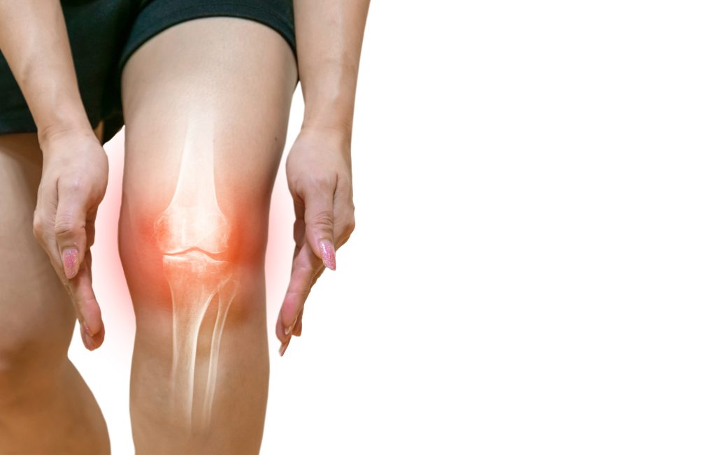
Joint cartilage when damaged, can lead to pain and even disability. There are several causes of joint cartilage damage, including physical damage, infection, osteoarthritis, rheumatoid arthritis, chemical damage, and infection. Understanding the causes of joint cartilage damage is essential for a good understanding of the condition. This helps in developing a personalized treatment plan and also keeping track of any changes in the condition.

What is Joint Cartilage?

Cartilage is a strong, organic and flexible tissue found throughout the body. It covers the surfaces of the joints between bones. It is an intermediate layer that reduces friction between bones and acts as a shock absorber. Type II collagen, which is a major structural protein in the human body is one of the components of the cartilage. Although cartilage is not bone, it does have cells called Chondrocytes just like the Osteocytes cells, found in bone tissue. Some types of cartilage are found at the ends of fingers and toes, in the ears, and in the nose.

Causes of Joint Cartilage Damage

Physical Damage

The causes of joint cartilage damage can be broadly grouped into physical and chemical damage. Physical damage to joint cartilage occurs when the cartilage is hit, scraped or injured by something like a bump or a fall. This might cause damage to the cartilage and also bring about a condition known as cartilage erosion, which leads to joint cartilage damage. In another form of physical damage to joint cartilage, the cartilage might be hit too severely by a hardobject like a ball or a bat, causing the tissue to bruise. This form of damage is reversible, but it still has an impact on the joint.

Chemical Damage

Joint cartilage is susceptible to chemical damage due to various reasons, such as an excessive amount of friction in the joint, certain medications, high fever, certain infections, and even certain types of surgeries. In the case of excessive friction, the cartilage can wear out, leading to joint damage. Excessive friction can also be due to a piece of bone or a joint capsule that is too thick, or that is out of place. If left untreated, this condition can lead to cartilage damage. Certain medications, such as non-steroidal anti-inflammatory drugs (NSAIDs) and even some types of birth control pills, can damage joint cartilage. High fever can also damage joint cartilage. This is because the body’s immune response to the infection causes an increase in joint fluid pressure, which damages the cartilage.

Infection

An infection in the joint cartilage can lead to damage to the tissue. Infections like bacterial and viral infections might spread due to injury to the joint cartilage in the form of a fall or a bump. It might also occur as a result of tissue impaction with foreign objects, like a piece of wood or even a splinter from your clothing. Bacterial infections are the most common type of infection that affects joint cartilage.

Osteoarthritis (OA)

Osteoarthritis is a condition that is caused by degeneration of the joint cartilage. It is specifically caused by aging, obesity, certain injuries, and even certain types of arthritis. OA is the most common cause of joint cartilage damage. It is one of the most common joint conditions seen in the elderly. On average, the condition affects two out of every ten people aged over sixty-five. OA is characterized by the progressive loss of joint cartilage, leading to abnormal bone growth on the cartilage surface. This abnormal bone growth leads to increased joint surface friction, resulting in pain and disability.

Rheumatoid Arthritis (RA)

Rheumatoid arthritis is an autoimmune dissorder that affects the joints. It is characterized by joint cartilage and bone inflammation, leading to joint damage and pain. People with this condition are likely to experience joint pain, swelling, stiffness, and even disability. Rheumatoid arthritis is the most common cause of joint cartilage damage. It is also one of the most difficult conditions to treat, as it usually does not respond well to medications. However, there are several surgeries available to help alleviate joint pain and disability.

Conclusion

While joint tissue exists everywhere in the human body, the extent to which it functions varies greatly between individuals. The function of joint cartilage depends on its structure and how tightly linked it is with other parts of your anatomy. When functioning properly, it provides stability to many joints within your body. For instance, moving your arm up and down repeatedly will place great stress on your elbow as well as other joints within that limb, helping to keep it stable while it undergoes these movements. An estimated 300 million people worldwide are affected by joint issues at some point in their lives. Hence, it becomes very important we look into the causes that can damage these joint cartilages as it provides stability, reduces the impact of movements, and helps protect the joint surfaces from damage. It is vital to your health and well-being, as it prevents your bones from rubbing together too much and causing damage.

Introduction

Artificial Intelligence (AI) has become an integral part in technological advancement in healthcare. AI significantly impacts many areas of the healthcare ecosystem, from prevention to diagnosis and treatment. Orthopedics, among all branches of medical specialties, is an area where surgeons have an array of new tools and assistants in the form of robots. Even though research is still around the success rate and superiority of robotic replacement surgery, the tech adoption has been very swift and global. Very soon, we will see exponential growth in robotic-assisted replacement surgeries that will outnumber manual knee replacement surgeries in a very short time. Patient concerns are mainly related to the procedure, recovery time, safety, costs, etc. 

What is Joint Replacement?

Joint replacement is a specialized procedure where artificial joints are implanted in patients with rheumatoid arthritis or osteoarthritis. It has been used as a solution for joint damage, especially in older people with osteoporosis and those who are injured frequently. This surgery is also effective in treating people with arthritis or arthritis associated with other joint diseases like polytraumatic arthritis or psoriasis. Some of the most common joints that are replaced include the hip, knee, and ankle joints. Joint replacement can be used to treat pain and restore function after a joint has been severely injured or due to a chronic joint disease such as rheumatoid arthritis or osteoarthritis. Artificial joints can be made from various materials, including metal alloys, plastic, and bio-implants like ceramic and polyethylene. This article will discuss the future of joint replacement and AI, why it’s important, and what can be done to improve it?

Impact of AI in Joint Replacement Surgery

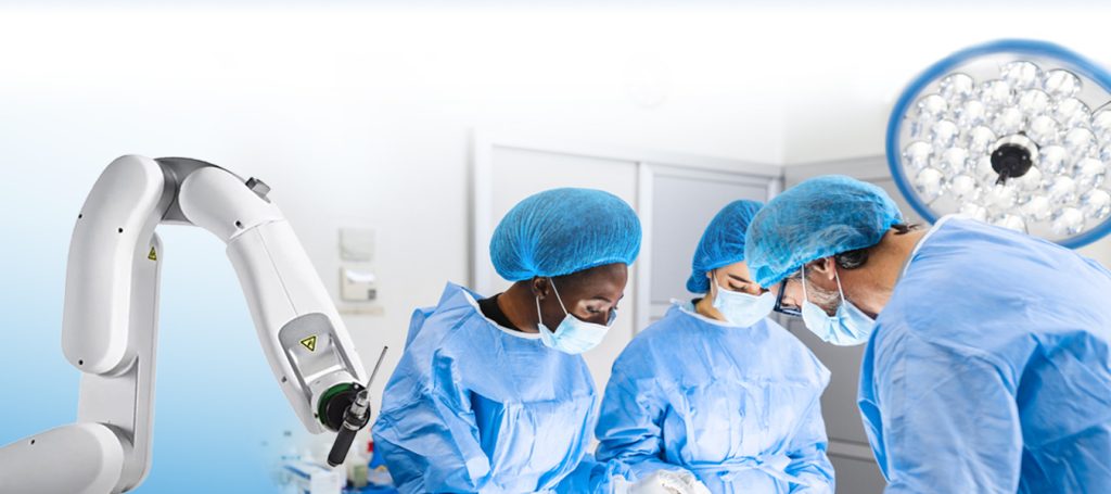
In joint replacement surgeries, robots typically assist with pre-surgery planning, intraoperative execution, monitoring and assessing complications, and any short-term or long-term outcomes. Robotic assistance in such surgeries can expand to using the computer vision model, sensors application, and feedback and loop execution, all of which will greatly help the surgeon for technical precision. 

Robotics with imaging is frequently used for 3D reconstruction, comparison of bone registration and ligament evaluation, performing bone resection, and collecting feedback. This leads to fewer mistakes and less damage to the soft tissues of the knee, as well as betters the implant alignment and corrects any flexion-extension gap and gap asymmetry. The fact that it predicts patient outcomes is an exciting prospect for both the patient and the surgeon. It promotes efficiency and faster healing times, improves precision and accuracy, personalizes patient care, and results in fewer complications. AI helps in surgical decision-making and eliminating risk factors and human-driven errors.

How can AI improve Joint Replacement?

AI can use genetic data to help select the best type of joint replacement for each patient. This would help reduce the number of patients who receive incorrect treatment, helping to improve the overall quality of care. AI can also be used to help doctors identify when a patient is at risk of developing a dangerous complication. This could include monitoring the health of a patient’s joint to identify any potential problem, like excessive wear on the joint.

The Future of Joint Replacement and AI

There is a constant effort to improvise the technology, learn from mistakes, and adapt to ensure that AI tools support surgeons with precision in their medical decisions and reduce human error. We are looking at a future with next-generation robots with advanced machine-learning algorithms capable of performing replacement surgeries with minimal human intervention. The impact of AI on the targeted segment will be huge as well. One area that can benefit from AI is joint replacement. 

Conclusion

Artificial joints/ Implants have been used for a long time now. The future of joint replacement, however, could be very interesting. AI could significantly improve the treatment of joint damage by automating a larger part of the process. This would allow doctors to spend more time on actual diagnosis and treatment, leaving less time wasted on administrative tasks. Patients who undergo joint replacement would be able to receive an AI-integrated treatment, like a virtual or augmented reality (VR/AR) treatment. AI could also be used to help select the right type of joint replacement for a patient based on their genetic profile. Researchers are focusing on newer technology for designing implants for  joint replacement with advanced and more biocompatible  material. The results of these studies can be helpful for the future of joint replacement.

Introduction

Joint replacement is the most preferred solution for the chronic joint pain accompanying arthritis and other degenerative joint conditions. But, as with any artificial joint, it comes with its challenges. In such a scenario, physiotherapy plays a significant role in the pre-operative and post-operative procedures in rehabilitating the muscle and the joints.

How is Physiotherapy Used Before Joint Replacement?

Physiotherapy can help you prepare for joint replacement surgery. Before a joint replacement, your doctor may recommend a program of joint exercises and physiotherapy to help you prepare your body for the change, including demonstrating what exercises should be done and how to properly execute them to reduce pain and any risk of injury. It is critical to focus on strengthening the muscles around the joint that is to be replaced. What that does is improve the chances of optimal recovery from surgery. An important thing to remember is that joint replacement surgery is not a way to cure arthritis; it is a way to relieve pain.

How is Physiotherapy Used After Joint Replacement?

In a joint replacement surgery, your doctor will remove all or part of your diseased joint and replace it with an artificial component. The material of artificial components is metal or plastic. These metal and plastic parts help your joint move freely or bend, making it more stable. You may need this surgery if your joints become worn out from overuse or if you suffer from a serious injury that damages your joints. After joint replacement, you’ll need to take it easy for a while to allow your body to heal. This means you’ll probably need to reduce the amount of activity you do physically. That, in turn, means you’ll need to turn to physiotherapy.

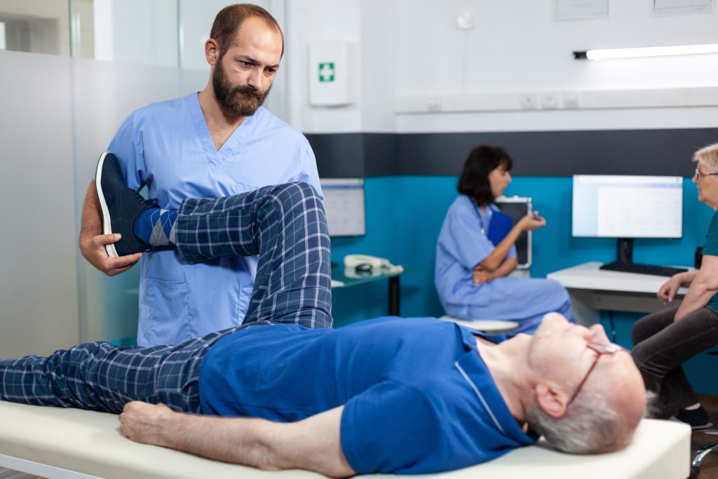
The post-surgery focus should primarily be on recovering the full range of motion and regaining full strength in the muscles around the knee. Physiotherapy at this point will include active exercises and stretches to improve the range of motion and gain muscle strength. The idea is to demonstrate and supervise strengthening programs to maximize recovery and administer modalities and manual therapy to minimize pain and swelling after the surgery.

The prognosis for recovery after Joint Replacement

Joint replacement is an effective way to relieve pain caused by arthritic conditions & trauma. Joint replacement also has the advantage of being a long term solution. The long-term prognosis for recovery after joint replacement is favorable, although the recovery process varies from person to person. For this reason, following your doctor’s post-surgery recovery plan is important.

Conclusion

As you can see, joint replacement aided with physiotherapy before and after the process has many benefits to offer. Physiotherapy can provide the physical therapy you need to recover from joint replacement surgery. Physical therapy can also help improve and restore movement after joint replacement surgery and prevent or manage some complications, if any. Physiotherapy can help manage chronic pain and swelling, help you get back to a normal lifestyle, and help you maintain or improve your quality of life. Physiotherapy also helps improve bone health, strength, and blood circulation, treat any accompanying pain and swelling, build strength in and around the joint, and aid in faster recovery. To recover as quickly and pain-free as possible after joint replacement surgery, following your doctor’s post-surgery recovery plan is essential.

Sinusitis – Symptoms, Causes, and Treatment

Sinusitis is an inflammation of the sinuses affecting the nose and occasionally the face, throat, and ears. Sinuses are hollow spaces in the skull behind the forehead, eyes, cheeks, and nasal passages. These sinuses get blocked and filled with fluid due to cold or allergies, resulting in inflammation. Sinuses are cavities in the skull, while sinusitis is a condition of inflamed sinuses.

How important are sinuses?

Sinuses play a key role in protecting the body against illness and taking care of our wellness. Sinuses, the hollow spaces filled with air, lighten the head’s overall weight. They also warm and humidify the air we inhale. Sinuses help enhance one’s voice quality and speech resonance. In an accident, the sinuses absorb the impact and protect the other facial structures. Sinuses also help drain nasal mucus that protects the nasal passage and keeps unwanted particles like dust and pollutants from entering the body. Healthy sinuses are empty except with a thin layer of mucus which continuously drains the germs, dust, and dirt out, keeping the nose clean and free from bacteria, as already mentioned above.

The four pairs of sinuses are-

Maxillary sinuses: Maxillary are the largest sinuses. They are positioned behind the cheekbones near the upper jaws.

Frontal sinuses: The frontal sinuses are divided into left and right frontal sinuses. The two sinuses are located at the centre of the forehead above each eye.

Sphenoid sinuses: Sphenoid sinuses are situated behind the eyes, near your optic nerve and pituitary gland.

Ethmoid sinuses: The Ethmoid sinuses are a collection of six to twelve small air pockets between the eyes and nasal bridge. 

One develops sinusitis when the tissue lining the sinuses get inflamed or swollen. Sinusitis can also be caused by viral, fungal, and bacterial infections. 

Symptoms of sinusitis

Sinusitis and common cold have similar symptoms, some of which may include the following-

Partial or total loss of smell called Anosmia

Fever, also known as Hyperthermia or pyrexia

Nasal congestion, which causes a stuffy or runny nose

Headache due to sinus

Cough or sore throat

Fatigue

As mentioned above, many sinusitis symptoms are similar to the common cold. But the main difference is that the symptoms of sinusitis tend to be more severe, last longer, and often include pain in the face and forehead. Sinusitis symptoms range from nasal congestion and facial pain to severe headaches and tooth pain.

Types of sinusitis

Depending on the severity and duration of the symptoms, sinusitis is grouped as –

Acute – Acute sinusitis, also known as Rhinosinusitis, where rhino means ‘the nose’ is the most common one with the shortest duration. The symptoms could remain for four weeks or less. Viral infections due to cold and seasonal allergies can cause symptoms that last up to ten days.

Subacute – In the case of Subacute sinusitis, the symptoms may last up to 12 weeks. Seasonal allergies or bacterial infections cause this type of sinusitis.

Recurrent acute – As the name suggests, there would be a reoccurrence of acute sinusitis at least four times in a year.

Chronic- Chronic sinusitis is when the symptoms last for at least 12 weeks.

Possible Contributors of Sinusitis

Anyone can develop sinusitis. Excessive mucus may get formed due to cold, bacteria, or allegories that gradually allow the bacteria and germs to grow in the sinuses, leading to infection. However, apart from cold, allergies, and viral or bacterial infection, other possible factors that may contribute to the development of sinus infection are-

  • Weakened immune system
  • Dental infection
  • Smoking and Tobacco
  • Structural issues with the nose like, nasal bone growth, uneven wall of tissue between the two nostrils (nasal septum)
  • Nasal polyps
  • Cystic fibrosis, a condition where thick and sticky mucus gets buildup in the lungs, intestine, and liver, which causes damage to the respiratory and digestive systems.
  • Exposure to mold, pollen, dirt, etc.

Treatment of Sinusitis

Sinusitis is curable and treatable in most cases without consulting a doctor and antibiotics. But if the symptoms of sinusitis persist for a long, it is advisable to see your doctor and follow his advice and medications. The doctor would diagnose sinus infection based on the symptoms and the physical exam. In case of chronic sinusitis, the doctor may recommend imaging tests like X-ray, CT scan or MRI. Blood tests, nasal endoscopy, and allergy tests are a few other tests that may be needed to diagnose the severity of the infection.

In many cases, home remedies or over-the-counter (OTC) pain relieving medicines would help treat headaches or pressure in the forehead or cheeks. The doctor will prescribe antibiotic therapy to block any sinuses’ growth in cases of bacterial infection. In cases of chronic symptoms that do not improve or heal with medication, sinus surgery would be an option to clear sinuses, treat structural deformity related to the nose or remove polyps. The types of sinus surgery are-

  • Functional Endoscopic Sinus Surgery (FESS)– In FESS, with the help of a magnifying endoscope, the surgeon views and removes the infected tissue or bone. With a small incision inside the nose, an endoscope equipped with a camera and light is inserted to help reduce the blockage and drainage of sinuses.
  • Turbinate Reduction Surgery – This surgery uses radio frequency to reduce the swelling of the turbinates (tiny structures in the nose that cleanse the air passing through the nostrils). A needle-like instrument is inserted into the swollen tissue to control the damage. The turbinates gradually heal and reduce in size allowing proper and easy breathing.

Sinus Ostial Dilation Surgery– In this type of surgery, with the help of a balloon catheter inserted into the blocked sinus passage, the balloon is inflated and a saline solution is injected to flush out the mucus.

Health-promoting lifestyle is one of the ways to prevent inflammation. This includes a healthy and nutritious diet, exercise, limiting exposure to allergens and irritants, maintaining good hygiene, and avoiding contact with infected people.

Knowing the causes and symptoms of sinusitis help in treating the inflammation timely and preventing the recurrence of the same in the future.