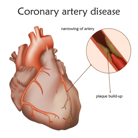
The possibility of having heart disease or a heart valve condition is overwhelming and frightening for most people. That is why having an understanding of heart diseases is important so that one can be well equipped with the right information, and hence take the right steps towards treatment.
Heart valve conditions are usually due to birth defects or age-related issues. Many heart valve conditions can be first identified with the help of a murmur, that is a whoosing sound as blood flows from one chamber to the next, or it may sound like an extra click when a valve allows backflow.
A murmur may indicate conditions such as Aortic stenosis, prolapse, regurgitation, etc. While medicines can be prescribed for a heart valve condition, they are mainly for the following reasons
- To reduce unpleasant symptoms from milder forms of the disorder.
- To maintain heart rhythm if a related arrhythmia is present.
- To lower the patient’s risk for clotting and stroke.
Valve diseases are generally progressive by nature, and the outlook for those who do not receive adequate and timely treatment could be fatal. If you are suffering from an aortic valve disease it is highly advisable to opt for a surgical procedure for low surgical risk patients.
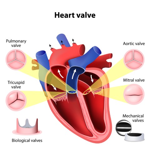
The heart has four chambers. The two upper chambers are called the left and right atrium and the lower chambers are called the left and right ventricle. The four valves that are at the exit of each chamber carry out the function of continuous blood flow through the heart to the lungs and the rest of the body. The four valves are the tricuspid valve, pulmonary valve, mitral valve, and aortic valve.
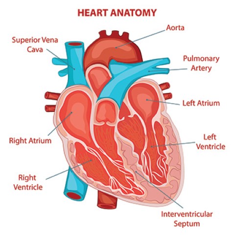
The aortic valve controls blood flow from the left ventricle into the aorta (the main artery in your body). When this valve opens, the oxygen-rich blood is pumped to the aorta and then to the rest of the body. It is key to the body’s blood circulation system.
Reasons you might need to undergo heart valve surgery
Types of aortic valve disease that may require treatment with aortic valve repair or replacement include the following:
Congenital heart disease: This condition may contribute to other complications that prevent the aortic valve from operating properly. For instance, a person may be born with a condition where the aortic valve does not have enough tissue flaps (cusps) or with a valve that is the wrong size or shape, or without an opening that allows blood to flow normally.
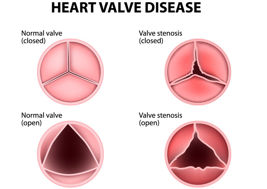
Aortic valve stenosis: When the aortic valve becomes narrow or obstructed, it becomes difficult for the heart to pump blood into the aorta. This may be caused by the thickening of the valve’s closure flaps, congenital heart disease or post-inflammatory changes associated with rheumatic heart disease. Around 75% of patients with unoperated aortic stenosis may die 3 years after the onset of symptoms.
Bicuspid aortic valve: A birth defect where only two cusps grow instead of the normal three. This is a common cause of aortic stenosis. Another cause may be that the valve opening does not grow as the heart does. This makes it harder for the heart to pump blood to the restricted opening. The defective valve becomes narrow and stiff over time because of a calcium build-up. Age-related Aortic Stenosis usually begins after age 60, but often doesn’t show symptoms until ages 70 or 80.
Aortic valve regurgitation: When the blood flows backward through the aortic valve into the left ventricle each time the ventricle relaxes rather than in the normal one-way direction from the ventricle to the aorta. This may be caused due to an abnormal valve shape present at birth (congenital heart disease) or by a bacterial infection.
Most symptomatic patients of AR will require valve replacement surgery within 2 to 3 years of developing breathlessness and symptoms of heart failure. A Swedish study has proven that these individuals have a life expectancy of 2 years lesser than the healthy population of the same age.
Valve diseases, although gradual and progressive are fatal if not treated in the right manner. Valve replacement surgery is recommended as it helps alleviate your symptoms and add quality years to your life.
Careful monitoring and doctor supervision may be all that is needed for a few people with mild aortic valve disease without symptoms. However, in most cases, aortic valve disease and dysfunction can get worse.
Such conditions eventually require surgery to reduce the risk of complications such as heart failure, heart attack, or a stroke or death by cardiac arrest. This is when a doctor will recommend heart surgery.
What are the consequences of not having a heart valve surgery?
- Decreased life expectancy and quality of life
As per an estimate, as the aortic valve disease gradually worsens, the average rate of survival without undergoing surgery is a mere 50% post 2 years, while it is 20% post 5 years.
While you may feel normal and not notice any problems for years, valve disease is not a condition to be ignored. Once you begin to experience symptoms like the ones mentioned above, not only does life expectancy and quality of life decline but the severity of diseases such as aortic heart stenosis increases.
- Contributes to more health complications
As the wall of the left ventricle works harder to pump blood through the narrow valve
opening into the aorta, the wall might show muscular thickening, along with the symptoms of aortic stenosis like fatigue and weakness.
The thickened wall allows less room for an adequate amount of blood circulated to the body, as it takes up more space inside the lower heart chamber. This may cause heart failure.
It is advisable to undergo valve replacement surgery as it not only adds to a better quality of life and solves the issue of aortic stenosis but also helps avoid the consequential conditions that come along with it.
Coping with Aortic Stenosis can not only be stressful physically but also mentally. Learning about your condition and the right treatment by talking to your doctor about it can help a great deal. Aortic Stenosis can be fatal and not undergoing the right treatment or prescribed surgery can lead to not just a deterioration in your condition but also create newer complicated conditions for your health. Knowing and understanding your condition as well as the benefits and success rates of the treatment gives you an upper hand and a better chance at survival.
Disclaimer: This blog is only for awareness purposes. We do not intent to promote any medications given in the blog. Please consult your physician before taking any medication.