Category

Surgery:

Category

Introduction

Any bodily abnormality may be genetic or caused due to injury, infection, exposure to radiation, or any reason that would affect a person’s appearance and, eventually, one’s self-esteem and sense of worthiness. As a medical specialty, plastic surgery covers a range of procedures that aim to improve a person’s appearance, repair and reconstruct bodily abnormalities and restore normal functions. Plastic surgery is a broad surgical field with two main areas: reconstructive surgery and cosmetic surgery. These surgeries often involve making incisions on the skin, which requires the use of sutures to close the wound and promote proper healing. Sutures are used to bring the edges of the skin together and hold them in place while the body naturally repairs itself. Various types of sutures are used in plastic and cosmetic surgeries, each with unique properties and benefits. Understanding the differences between these sutures is crucial for surgeons to choose the appropriate type for each patient and to achieve the best aesthetic outcome.

This article will discuss some of the most commonly used sutures in plastic and cosmetic surgeries and their advantages.

What is Cosmetic Surgery?

Surgery that focuses on improving and enhancing a person’s physical appearance is known as cosmetic surgery. Cosmetic surgery is performed on people who are otherwise healthy and want to improve or enhance their appearance for personal, aesthetic reasons.

The face, breasts, abdomen, arms, and legs are just a few of the body parts that might undergo cosmetic surgery. Breast augmentation, liposuction, rhinoplasty, facelifts, and stomach tucks are a few of the most popular cosmetic surgery operations.

Each patient’s personal goals and preferences are considered during the highly individualised cosmetic surgery procedure. Patients typically speak with the surgeon before a cosmetic operation to go over their expectations, goals, and any possible issues. The surgeon will then evaluate the patient’s health and suggest the best course of action.

Cosmetic surgery entails risks and potential problems, just like any other form. To pick a trained and experienced surgeon who can carry out the treatment safely and successfully, patients should carefully weigh the risks and benefits of the procedure.

What is Reconstructive Surgery?

Reconstructive surgery is performed to repair the abnormality of the person’s body part. This type of surgery mainly aims to treat any such abnormality, whether caused by birth or due to injury, disease, age, or any other reason and restore the normal appearance and function of such body parts.  

The discipline of plastic surgery is intricate and complex, requiring a lot of training and specific knowledge. Plastic surgeons must thoroughly understand anatomy, wound healing, and several surgical procedures. There are many various kinds of plastic surgery operations, such as liposuction, facelifts, rhinoplasty, and breast augmentation. Every procedure is carried out with the utmost care and precision and customized to the specific patient’s demands and goals.

It is important to remember that cosmetic and plastic surgery is different. Despite their frequent confusion, reconstructive and cosmetic operations are included in the broader field of plastic surgery. While cosmetic surgery seeks to improve the appearance of a generally healthy person, reconstructive surgery aims to restore function and normal appearance following an injury or sickness. 

Plastic surgery’s main aim is the reconstruction of facial and bodily defects and, by nature, is reconstructive. 

What are commonly used sutures in plastic and cosmetic surgery?

An ideal suture must cause the least injury to the tissue and skin, the least tissue reaction, one that is easy to handle, has high tensile strength, provides proper knot security, and can be used in any tissue. However, factors such as the wound type, infection risk, the healing time required, suture properties, patient’s skin type, age, and genetic history decide the selection of sutures in plastic and cosmetic surgery. 

Every single plastic and cosmetic surgery procedure has its own set of sutures, depending on the operation and the result expected from the surgery.

Titanium Sutures

Titanium suture is made of titanium alloy wire. It is strong yet has a low rate of infection. It is used alongside other less invasive sutures, like polyglactic catgut or polyglycolic acid. It is non-absorbable, which means they are not absorbed by the body. These sutures do not leave scars, so they are ideal for many cosmetic procedures like face lifts, augmentation surgeries, tummy tucks, and hair transplants. However, it is not recommended for liposuction procedures.

Chromic Catgut Sutures

Of all the sutures commonly used in plastic or cosmetic surgery procedures, chromic catgut suture is the strongest, made of natural fibre. It is highly absorbable, which is ideal for reconstructive surgeries. It can be used in many body parts like the face, hands, feet, and joints. It is flexible and pliable with high tensile strength, which makes it perfect for procedures like hand revisions, joint revision surgeries, and facial surgeries like brow lifts and facelifts. Chromic catgut sutures are available in both natural and black colours. The natural variety is more suitable for lighter skin tones, while the black one is ideal for darker skin tones.

Polyglactin Sutures

The most popular alternative for general soft tissue approximation is the Polyglactin Suture, made of a synthetic braid and suitable for treating wounds on the hands and face. This suture is also utilised during vascular anastomosis procedures, just like the Poliglecaprone suture. In contrast to catgut sutures, polyglactin sutures have a more predictable absorption level and usually cause a modest tissue reaction for the duration of the absorption process. Moreover, there is little to no tissue reactivity to this suture.

Monofilament suture: nylon and prolene

These sutures are made from synthetic fibre and are non-absorbable and strong. They are used for reconstructive surgeries like hand and foot surgeries, shoulder repairs, and facial procedures like brow lifts, blepharoplasties, and jawline revisions. Monofilament sutures are available in various colours, like green, blue, and red. Moreover, they are suitable for all skin types, making them an ideal choice for cosmetic surgery. 

Metallic threads: silver, gold, and platinum

These threads are metal and ideal for ear tuck surgeries and nose reshaping. However, they are not suitable for liposuction procedures. They are the most expensive sutures and must be cared for carefully by the surgeon.

Silk Fibre Suture: Cohesive and Non-cohesive

Silk fibre sutures are made from the filament of a silkworm. They are an ideal option for cosmetic surgeries like facelifts and laser resurfacing. They are also suitable for reconstructive surgeries like hand reconstruction and joint replacement. They can be used in a variety of body parts like hands, feet, face, ears, and lips. 

Short bowstring sutures

In this suture, a short string made of silk is tied around the base of the needle. The needle is then inserted into the body part, which makes this suture quite similar to a monofilament suture. However, it is available in a shorter length of 5mm compared to the 9mm of a monofilament suture. It is strong, flexible, and pliable, which makes it an ideal option for cosmetic surgeries such as eyelid surgery, facelifts, blepharoplasties, and body contouring surgeries.

Bottomline

There is no denying that plastic and cosmetic surgeries can change your life for the better. If you are thinking of getting one done, make sure you research thoroughly and find a qualified surgeon. Many types of sutures are used in plastic and cosmetic surgeries, each with advantages and disadvantages. 

Introduction

Ophthalmology is a branch of medicine that deals with diagnosing and treating eye disorders. The use of laser technology in ophthalmic surgery has witnessed a significant advancement in recent years. Suture, one of the oldest and most popular methods of ligation in ophthalmic surgery, has been falling out of favour with the introduction of laser-assisted ligation (LAL) techniques. Many ophthalmic surgeons view sutures as an ‘aging method’ that is not useful in surgical precision. However, this may no longer be the case, as new research shows new uses for sutures that make them relevant again in ophthalmic surgery. Despite cataract surgeries being performed without sutures, suturing is still an essential skill that every eye surgeon must master well. To understand why this is the case, we must look at what suture is and how it works. Part of this skill requirement arises from knowing and deciding which suture to use. In this blog post, we will explore why suture is still relevant in ophthalmic surgery, even in the age of lasers.

What is a suture?

Suturing is a method of tying off blood vessels to prevent bleeding and hold a wound together till the natural healing process is sufficiently established. It consists of a thin, long thread that is twisted into a loop and either sewn into the tissue or tied with a knot. The introduction of sutures in ophthalmic surgery dates back to the mid-1800s when it was used to stop bleeding after surgery to the blood vessels. A suture is commonly used in ophthalmic surgery to close incisions or wounds. Since the eye is a delicate organ, sutures provide the necessary support to the surrounding tissue. It helps keep the wound closed and prevents scar tissue formation. This is especially important in the case of cataract surgery, which is the most common eye surgery performed worldwide.

Why Does Suture Matter in Ophthalmic Surgery? 

Laser technology has revolutionized the field of ophthalmology, but it has its limitations. Some ocular conditions and anatomies may not be amenable to laser surgery and therefore require traditional suture techniques. For example, laser surgery is not suitable for certain types of retinal detachment, and a suture is used to repair the detachment. Therefore, sutures can be used in conjunction with laser surgery.

As already mentioned above, laser technology may not be for everyone. 

  • Factors like family history, medical conditions, and lifestyles make some people non-eligible for laser treatment.
  • People with autoimmune diseases, with an inability of the body to heal or prone to infections, are not eligible for laser treatments, i.e., diabetic patients who take longer to heal or people with high blood pressure and hormone imbalance. 
  • Laser treatment may cause dry eyes. 
  • In case of cataracts, a small incision is becoming common. However, for people with dense lenses, the traditional procedure called the Extracapsular Cataract Extraction (EECE) is advisable.
  • Age is also one of the deciding factors. Patients below 18 years are not good candidates for laser treatment as their vision changes over time, and the laser is permanent.
  • Those taking certain medications may experience the side effects of laser treatment.

How do Sutures work in Ophthalmic surgery?

Suture plays a crucial role in closing incisions, supporting the surrounding tissue, and in some instances, providing additional stability and strength to the repair. With the advancement in suture materials, using sutures in ophthalmic surgery is becoming more comfortable and less invasive for patients. Sutures will continue to be an essential tool for ophthalmic surgeons for many years.

In addition, the suture is often used with other technologies, such as laser surgery, to provide the best outcome for the patient. For example, a laser may be used to make an incision, and then a suture is used to close it. This allows the surgeon to take advantage of the precision and control of sutures while also benefitting from the benefits of laser technology.

Advantages of Suture in Ophthalmic Surgery 

The advantages of sutures in ophthalmic surgery are many. One of the key advantages of sutures in ophthalmic surgery is their versatility. The suture can be used in various procedures, from simple incision closures to complex reconstructions. Suture takes less time to tie than laser. For example, sutures can be used to repair a detached retina, to close a wound after a corneal transplant, or to reattach a detached iris. It is safe for eyes with cataracts, glaucoma, etc. 

Another advantage of sutures is their precision. Suture allows the surgeon to make tiny and precise incisions, which is especially important in the delicate structures of the eye. The suture also allows the surgeon to control the tension on the wound, which is crucial for proper healing. 

The suture is also a reliable and long-established technique used for many years in ophthalmic surgery. Surgeons are highly trained and experienced in suture techniques, which means that the risk of complications is low. Furthermore, the materials used for suturing have also improved over time, becoming stronger, more flexible, and less likely to cause irritation. Suturing does not cause pain or inflammation. 

It’s also important to note that suturing is a cost-effective option for patients as it does not require special equipment. Suture materials are relatively inexpensive, and the procedure is typically less expensive than laser surgery. Furthermore, the suture is also a good option for patients in remote or underserved areas, where access to advanced technology may be limited.

In addition, sutures can provide additional stability and strength to the repair in certain cases. For example, the suture is used in corneal transplantation surgery to hold the transplanted tissue in place. The suture also helps to prevent the transplanted tissue from being dislocated. Sutures can be used on eyes with very weak or no muscle function.

It’s important to note that the use of sutures in ophthalmic surgery has also improved over the years. With technological advancement, suture materials have become stronger, more flexible, and less likely to cause irritation or allergic reactions. Dissolvable sutures are also becoming more common, which eliminates the need for suture removal and leaves no scar tissue.

Conclusion

In conclusion, while laser technology has revolutionized the field of ophthalmology, the suture is still an essential tool in ophthalmic surgery. The suture is still relevant in ophthalmic surgery in the age of lasers because of its versatility, precision, reliability, and cost-effectiveness. While laser technology has revolutionized the field of ophthalmology, suture remains an essential tool for ophthalmic surgeons to provide the best outcome for their patients.

In case you are a caregiver of a patient who is scheduled for ophthalmic surgery, it’s important to understand the suture’s role in the procedure. It’s also important to ask your surgeon any questions you may have about the use of sutures in surgery. Remember, an informed patient is a more confident patient.

FAQs

Q: Is suture used in all ophthalmic surgeries?

A: No, sutures are not used in all ophthalmic surgeries. Some procedures, such as laser surgery for cataracts, do not require sutures. However, the suture is commonly used in many ophthalmic surgeries, such as traditional cataract surgery, retinal detachment repair, and corneal transplantation.

Q: Does suturing hurt?

A: The use of sutures during surgery is typically done under local or general anaesthesia, so the patient should not feel any pain during the procedure. After the surgery, there may be some discomfort or pain, but this is usually mild and can be managed with medication.

Q: How long does it take for sutures to heal?

A: The healing time for sutures varies depending on the type of surgery and the patient’s immunity and response to the healing process. The sutures are generally removed within a few days to a few weeks after the surgery. The body will absorb dissolvable sutures over time. Your doctor will give you detailed information about the healing process.

Q: Will I have a scar after the surgery?

A: Incisions made during ophthalmic surgery are typically tiny and are placed in areas that are less visible to the naked human eye. This means that the scarring is usually minimal and not noticeable. However, it’s important to keep the incision area sterile and clean and follow your surgeon’s post-operative instructions to minimize scarring.

Q: Are there any risks associated with sutures?

A: As with any surgical procedure, there are risks associated with using sutures. However, these risks are generally low, and most patients experience no complications. Some risks include infection, bleeding, and allergic reaction to the suture materials. It would help if you inform your surgeon about your allergy history, and they will discuss these risks before the surgery.

Q: Can I resume my normal activities after the surgery?

A: The recovery time from an ophthalmic surgery varies depending on the type of surgery and the patient’s healing process. Your surgeon will give you specific instructions on when to return to work and normal activities. Usually, you will need to avoid any heavy-duty impactful, or strenuous activities and keep your eye protected for a few weeks after the surgery.

Introduction

Coronary artery disease (CAD) is a serious condition that affects millions of people around the world. It is a form of cardiovascular disease that is caused by the hardening and narrowing of the arteries that supply blood to the heart. If left untreated, ‍coronary artery disease (CAD) lead to chest pain, complete blockage of an artery, heart attack, and cause sudden death. Knowing the symptoms, risk factors, and treatments for CAD can help you protect yourself and those around you from its potentially devastating effects. In this article, we’ll explore what you need to know about coronary artery disease and how to identify and manage this devastating condition and keep it at bay. However, there are also various preventative measures that you can take to reduce your risk of developing the condition. With the right information and knowledge, you can reduce your risk of developing this dangerous condition and improve your overall heart health.

What is coronary artery disease?

Coronary artery disease (CAD) is a medical condition in which plaque builds up in the arteries that supply blood to your heart.  The buildup of fatty deposits in the arteries is a common precursor to CAD, this build-up of fatty deposits in the arteries could be formed over a period of time, which can completely block the arteries and cause heart failure. This buildup causes the arteries to narrow, which increases your risk of a heart attack. The plaque that builds up in the arteries can be made of cholesterol, calcium, and pieces of fatty material. It usually starts in the walls of the arteries, and it can build up and grow bigger over time, which puts pressure on the artery walls. This can make it harder for your heart to get the blood it needs. In most cases, CAD is the result of a combination of risk factors that increase your chances of developing it. These risk factors include aging, smoking, high blood pressure, high cholesterol, diabetes, and obesity. While these risk factors can be controlled with lifestyle changes, there are treatments that can help reduce your risk of developing CAD.

Symptoms of coronary artery disease

The first and most common symptom of CAD is chest pain. The pain may feel like pressure, squeezing, or an achy and heavy feeling in your chest. It can be mild or severe, and it can feel like something is pressing on your chest or that it’s hard to breathe. Other symptoms of CAD can include

  • shortness of breath,
  • weakness or fatigue,
  • dizziness or lightheadedness
  • palpitations,
  • pain or pressure in your shoulders or arms,
  • nausea or vomiting,
  • sweating,
  • fainting.

If you experience any of these symptoms, it’s important to see your doctor. The sooner you get treatment, the better your chances are of preventing a heart attack and reducing your risk of mortality.

Risk factors of coronary artery disease

As mentioned above, most cases of CAD are caused by a combination of risk factors. These factors may include aging, smoking, high blood pressure, high cholesterol, diabetes, and obesity. Depending on your individual risk level, these risk factors can lead to CAD in two ways: They can cause the buildup of plaque in the arteries, or they can increase your risk of blood clots forming in the arteries.

Diagnosis of coronary artery disease

If your doctor suspects that you have CAD, they would advise on heart health evaluation tests to diagnose further. This includes a thorough history of your symptoms and a physical examination. They may start with a blood test to measure your cholesterol levels and assess the extent of your risk for CAD. If your cholesterol levels are high, you may need to take medication to lower them. Your doctor may also order a blood test to look for signs of diabetes. They may also recommend an electrocardiogram (also known as an EKG or ECG) to check for signs of a heart attack or an abnormal heart rhythm. If your doctor suspects that you have CAD, they may recommend a coronary artery calcium test. This test uses X-rays to scan your arteries and look for signs of plaque. If you have a high amount of plaque in your arteries, it could mean that you have CAD.

Treatment of coronary artery disease

If you have established CAD, your doctor will work with you to develop a treatment plan. Depending on your specific case, they may recommend lifestyle changes, nutrition support, medication, or even surgery. In certain cases, doctors may even recommend a procedure called a percutaneous coronary intervention (PCI). This minimally invasive procedure can help you reduce your risk of heart attack by removing plaque from your coronary arteries. The treatment options include interventional procedures and treatments with stents, coronary artery bypass grafts (CABG) or medications depending upon the case. Stents are small mesh tubes that are inserted into the arteries. Medications can help lower your cholesterol levels, blood pressure, and blood sugar. CABG is a surgical procedure that reroutes blood flow around blocked arteries.

Prevention of coronary artery disease

The best way to prevent CAD is to initiate self-care and maintain a healthy lifestyle. This means eating a balanced diet, getting enough exercise, and maintaining a healthy weight. A healthy diet and an active exercise routine will help you in your journey to maintain a healthy weight and improve your heart health. A healthy diet should consist of various fruits, vegetables, lean proteins, whole grains, low-fat dairy products, and salt-free table salt.

Make sure to maintain healthy blood pressure and cholesterol level and a moderate body mass index (BMI, a measurement of body fat based on height and weight). These will help prevent CAD and reduce your chances of a heart attack or stroke. The best way to prevent CAD is by managing your risk factors. This means taking steps to reduce your risk of developing each of the risk factors for CAD. With the right lifestyle changes, you can greatly reduce your risk of CAD and heart disease. Some of the most important lifestyle changes include eating a healthy diet, getting regular exercise, and managing your stress levels. Your doctor would prescribe certain medications for controlling cholesterol, reducing chances of blood clots formation, and maintaining healthy blood pressure, if need be. If you’re concerned about your risk of developing CAD, it’s important to visit your doctor and have them perform a full checkup. During your appointment, they’ll ask you about your medical history, lifestyle, and family history. They’ll also perform a physical exam and order any necessary tests to help you get a better understanding of your current heart health. Based on the results of your tests, your doctor may recommend lifestyle changes, medication, or even surgery to reduce your risk of CAD.

Tips for improving coronary artery health

If you’re concerned about your risk of developing CAD, there are several things you can do to protect your heart. Make sure you’re getting enough exercise. The best exercise is one that you enjoy and that you can do regularly. It’s also important to make sure you’re getting enough sleep, managing your stress levels, and following a healthy diet. To help prevent CAD and improve your heart health, follow these tips:

  • Stay away from tobacco products.
  • Maintain a healthy weight, and eat a balanced diet that’s rich in fruits and vegetables.
  • Get your cholesterol and blood pressure checked regularly.
  • Keep your diabetes under control with regular exercise and a healthy diet.
  • Visit your doctor regularly and get your blood pressure checked.

Conclusion

Knowing the symptoms, risk factors, and treatments for CAD can help you protect yourself and those around you from its potentially devastating effects. In this article, we’ve explored what you need to know about coronary artery disease and how to identify and manage it. We’ve also discussed aspects of treatment options and preventive measures. If you have any of the above-mentioned CAD risk factors, it is essential to be evaluated for CAD, even if you do not experience symptoms. Following a healthy diet and exercise routine is essential to maintain a healthy heart.

FAQs

At what age do your arteries start clogging?

Research has shown that on an average the from the age of 35 and upwards, most men and women start to have blocked arteries. 

What are the signs that your heart is weak?

Signs like shortness of breath on exertion, chest pain, fatigue, and swelling of the legs, ankles, and feet indicate the heart is weak.

What does a blocked artery feel like?

A blocked artery will induce symptoms of chest pain and tightness, and shortness of breath.

Can an ECG detect a blocked artery?

An ECG can detect a blocked artery. Also, your cardiologist may recommend an ultrasound to check for blockages

Is coronary artery disease and heart disease same?

Cardiovascular disease or heart disease refers to a cluster of diseases related to the heart, including coronary heart disease.

Does coronary artery disease require surgery?

If the arteries are blocked in several areas, or if there is a blockage in one of the larger main arteries, then coronary bypass surgery will be recommended.

Can a blocked artery clear itself?

No, a blocked artery does not clear itself.

What is the test recommended for coronary artery disease?

Coronary Angiography, also called cardiac catheterization, is the recommended test for diagnosing coronary artery disease.

Introduction

Blood vessels responsible for transporting blood throughout the body are the components of our circulatory system. Arteries, veins, and capillaries are the various blood vessels, each performing a specific function to regulate blood circulation, ensure the proper supply of oxygen and nutrients, and removal of waste from the body. Arteries supply oxygenated blood from the heart to different parts of the body, while veins return the deoxygenated blood from the rest of the body to the Heart. The capillaries act as a connection between the arteries and the veins.

Any blockage affecting the smooth blood flow in these blood vessels leads to various health problems relating to the heart, brain, and other body parts. Blood circulation disorder affecting the blood vessels and blood supply to –

  • The heart is called Coronary Artery Disease
  • The brain is called Cerebrovascular Disease
  • The body parts other than the heart and the brain is called Peripheral Vascular Disease.

In this article, we will discuss Peripheral Vascular Disease and know the causes, symptoms, types, and treatment of the diseases.

What is Peripheral Vascular Disease?

Peripheral Vascular Disease is the condition of blockage and narrowing of the arteries and veins in the legs, arms, and body parts other than the heart and the brain. Peripheral Artery Disease (PAD) and Peripheral Vascular Disease (PVD) are often used interchangeably. However, there is a difference between the two. PAD is the narrowing of the arteries due to the accumulation of plaque (i.e., fats and cholesterol) on the inner walls of the arteries that impair the blood flow from the heart to different parts of body, which remain deprived of oxygen and nutrients. While PVD refers to the blockage and narrowing of the arteries and veins in parts of the body other than the heart and the brain due to various reasons, including the accumulation of plaque. Thus, PVD is an umbrella term.

Causes of PVD

The primary cause of PVD is Atherosclerosis. The build-up of plaque, i.e., accumulation of fats and cholesterol, can be in any artery in the body. The fatty substances and cholesterol that block the arteries and thereby reduce the blood flow are known as Atheroma. The condition of this plaque formation in the arteries is called Atherosclerosis. Due to narrowing, when the arteries cannot supply enough blood to the legs, it causes pain, cramps, aches, and discomfort during physical activity, but the same subsides with rest. This condition of pain in the legs is called claudication. Atherosclerosis can happen in any body part. But it may be noted that Atherosclerosis is caused in the arteries and not in the veins.   

Besides, Atherosclerosis, the other responsible factors for PVD can be-

  • Coronary Artery Disease – people with blocked arteries of the heart are at risk of developing PVD.
  • Lifestyle – Lifestyle choices like smoking, sedentary living, poor eating habits, and drug use also play a significant role in developing PVD.
  • Medical conditions– Medical conditions include diabetes, high blood pressure, high cholesterol, obesity, and kidney disease, which may also be the reason for one developing PVD.
  • Injury – Injury to the legs and arms may cause the veins to narrow or swell, impairing blood flow from the legs or arms to the heart.

Symptoms of PVD

People with PVD may not experience any symptoms in the early stages. However, the symptoms depend on the blood vessels blocked and the extent to which they are blocked. Some of the symptoms that people affected with PVD may experience are –

  • Pain with physical activity, i.e., cramps, aches in the hip, buttocks, calf, and thigh which subside with rest.
  • Weakness in muscles
  • Shiny and smooth skin
  • Swelling, sores in legs that do not heal
  • Numbness and coldness in toes, feet
  • Reduction of pulses in feet
  • Decolouration of the skin
  • Severe burning
  • Loss of hair in the affected area

Types of PVD

Depending upon the nature of damage caused to the blood vessels, PVD is categorized as Occlusive and Functional.

  • Occlusive PVD

When there is physical damage to the vessels, resulting in structural changes in the blood vessels, it is the case of occlusive PVD. The following are examples of occlusive PVD-

Atherosclerosis– When the walls of the arteries thicken or harden over a period due to the accumulation of plaque, obstructing the smooth flow of blood in the arteries.

Carotid Artery Disease– When the large arteries (carotid arteries) on either side of the neck carrying oxygenated blood to the brain are narrowed. It is known as Carotid Artery disease or Carotid Stenosis. 

Lymphedema– Lymphedema is a condition where the fluid in the lymphatic system builds up. The lymphatic system is the network of thin tubes and lymph nodes (small structures that filter harmful substances) running throughout the body to help eliminate body toxins and waste, fight against infection, and fuel the immune system. The body parts of the affected lymph nodes get swollen.

Abdominal Aortic Aneurysm – The part of the Aorta (the largest artery supplying oxygen-rich blood to all the body parts) that runs through the abdomen is called the abdominal Aorta. Aneurysm refers to the outer bulging (like a balloon or a bubble) on the blood vessel. This is caused due to weakening of the artery walls. An aneurysm may occur on the abdominal Aorta, which can be life-threatening with internal bleeding if there is a rupture in the artery. 

Deep Vein Thrombosis (DVT) – DVT is when a blood clot is formed in the deep vein, often in the leg, arm, or pelvis, resulting in swelling in the veins, cramping, and redness in the affected area. If the clot breaks, its fragments can travel to any body part.

Buerger’s Disease– This is chronic inflammation in the small arteries of the extremities (the hands or the feet), causing a blood clot and blockages in the legs or arms.

Popliteal Artery Entrapment Syndrome (PAES) – PAES is a rare vascular condition where the muscles and tendons near the knee compress the popliteal artery (the main artery behind the knee), thereby restricting blood flow to the lower part of the legs. This usually is common among athletes.

A few other diseases caused due to blockage of arteries leading to the particular organ of the body are Intestinal Ischemic Syndrome which affects the gastrointestinal system, and RenalArtery Diseasecausing damage to the arteries supplying blood to kidneys .The term Ischemic refers to the lack of blood supply to a body part that deprives it of oxygen and nutrients.

  • Functional PVD

When factors such as stress, cold atmosphere, smoking, or vibrating of machinery the blood vessels to widen or narrow, it is the case of Functional PVD. There is no physical damage or structural change in the blood vessels here.

Raynaud’s Disease– Exposure to cold temperature, anxiety, and emotional disturbances cause Raynaud’s disease, in which the blood flow to the fingers, toes, ears, etc., is impaired.

Chronic Venous Insufficiency (CVI) – CVI is a condition when the valves of the veins do not function properly and hence cannot transport blood back to the heart. The blood flows back to the veins instead of the heart, and this causes swelling or aching in the legs or feet. Varicose veins are twisted and enlarged veins visible under the skin caused by venous insufficiency.

Treatment of PVD

PVD affects cardiovascular health, and there is a high risk of life-threatening complications of heart attack and stroke if left untreated.

Healthy living with positive lifestyle changes like adequate physical activity, a proper nourishing diet low in fats, and smoking cessation can help avoid and reduce the risks of PVD.  

Timely treatment is of utmost importance in reducing the severity of PVD. The treatment starts with your doctor recommending various tests like-

  • Ankle Brachial Test, which measures the blood pressure at the ankle or the arms when the patient is at rest, or
  • A treadmill test is conducted to check the pain threshold while the patient walks on the treadmill.
  • Ultrasound Doppler to check the blood circulation in the arteries of the legs to ensure the presence of any blockage therein. 
  • Computed tomography angiography (CTA) and Magnetic resonance angiography (MRA) tests are carried out to detect clogged/narrowed blood vessels. 
  • Angiography- Angiography is conducted with the help of dyes that are injected into the arteries to detect the blockages.

Depending on the need and the severity of the condition, your doctor would suggest medications only or an interventional procedure with medications to treat PVD. 

Peripheral Angioplasty is a minimally invasive procedure performed under local anesthesia to open the clogged artery and restore normal blood flow. A diagnostic catheter (a thin tube) is inserted through a tiny incision in the thigh. With the help of a Live X-ray, the operator can guide this catheter to the site of the blockage in the artery. A balloon is inflated inside the artery to press the plaque to the walls of the artery, making way for normal blood flow. A stent support may also be provided to the treated artery to keep it open. Once the stent is placed correctly, the incision is closed.

Conclusion

PVD is a slow and gradually increasing circulatory disorder. The symptoms are not noticeable at the early stages. However, those experiencing the symptoms must consult a doctor for timely and proper diagnosis and treatment. This is so because the major risks associated with PVD are heart attack or stroke complications which can be life-threatening as already mentioned above. In severe cases, this may also result in tissue death leading to limb amputation

Always follow a healthy lifestyle, and ensure timely and appropriate medical intervention and treatment to avoid and reduce the risks of Peripheral Vascular Diseases.

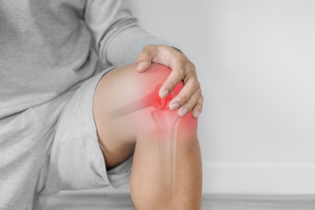
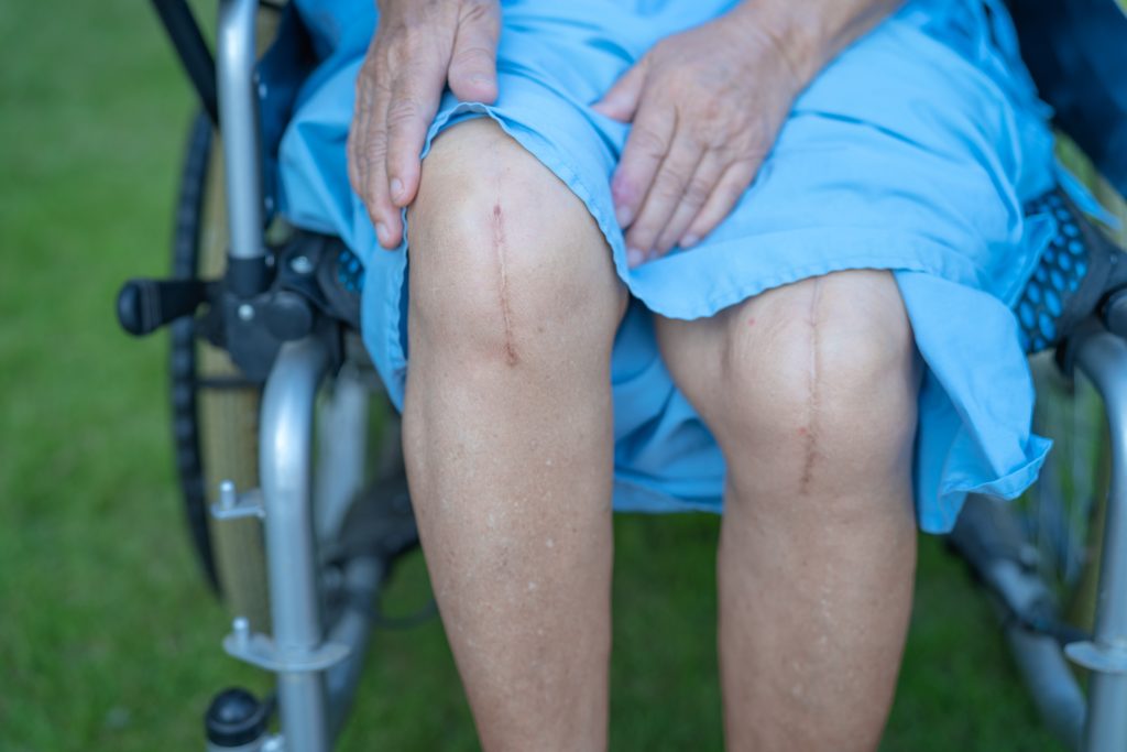
Knee replacement surgery is a common and effective way to restore mobility and relieve pain in patients with degenerative arthritis of the knee. In most cases, knee replacement surgery is successful and produces excellent results. The procedure restores much of the original functionality of your knee joint while also addressing any underlying causes of pain and disability. However, the very idea of having to undergo surgery creates stress and anxiety in many people, especially the elderly, which is quite normal and understandable. In such an event, immediate family members and caregivers need to step in and do what it takes to prepare them for the upcoming surgery. 

What is a knee replacement?

A knee replacement is a procedure in which a surgeon replaces your knee joint with an artificial joint. The artificial joint is made of a metal and plastic component that is surgically implanted. Knee replacement surgery treats people who have advanced osteoarthritis or rheumatoid arthritis. Knee replacement surgery restores function to the knee joint. You may notice an increased range of motion and decreased pain after knee replacement surgery. 

How to Prepare Seniors for Knee Replacement Surgery

Preparing our elderly folks for surgery is one big factor in the success of the surgery. The surgery’s run-up is critical to success, healing, and recovery. Knee replacement surgery is a major procedure that can significantly improve mobility and reduce pain for elderly individuals with severe knee damage or degeneration. Preparing for knee replacement surgery can be a challenging process, especially for elderly individuals who may have additional health concerns or mobility issues. 

The role of caretakers includes:

  • Taking care of the patient’s emotional health.
  • Sharing success stories of other patients to boost their morale.
  • Ensuring their good health and diet before surgery.
  • Providing moral support.
  • Creating an environment of acceptance and readiness to help them come through successfully.

Here are some steps you can take to help prepare your elderly loved one for knee replacement surgery:

  • Help them understand the surgery: Make sure they understand the surgery well and what to expect before, during, and after the procedure. Of course, the surgeon and their team will brief the patient and their immediate family about the surgical procedure, what it entails and the outcome, and all the nitty-gritty. However, as a caregiver, it is your responsibility to try and comprehend the medical implications of this surgery and pass on the net positive information to the patient. This hand-holding will also reduce the fear, stress, and nervousness that come before undergoing a surgical procedure. 
  •  Follow the surgeon’s instructions: The surgeon will provide specific instructions on preparing for the surgery, such as fasting for a certain period before the procedure. It’s also important for the elderly to follow their surgeon’s instructions and attend any pre-surgery appointments or educational sessions. By preparing in advance and working closely with their healthcare team, they can increase their chances of a successful surgery and recovery.
  • Discuss medications and allergies: Make sure to inform the surgeon and anesthesiologist about any medications your loved one is currently taking and any allergies they may have.
  • Do Your Research: Additionally, reading up on doctor-backed articles and information about the procedure for the layperson is helpful. Do make sure all doubts and questions are addressed before the surgery. Constant reassurance to your elderly loved one that you will be there before, during, and after the surgery. 
  • Attend pre-surgery tasks and appointments: Your loved one must attend all pre-surgery appointments with their surgeon and anesthesiologist to discuss the procedure and any potential risks or complications. Help them gather necessary medical documents or reports, fill out paperwork, and arrange transportation to and from the hospital.
  • Prepare the home: Consider making any necessary adjustments to the home to make it easier for your loved one to move around after the surgery and make the recovery process more manageable. This may include installing handrails, removing tripping hazards, and ensuring all necessary items are within easy reach. This assures them that their needs and comfort are taken care of and acts as a morale booster.
  • Help them get in shape: Encourage them to engage in gentle exercises to strengthen their muscles and improve their overall physical condition before surgery.
  • Make arrangements for aftercare and recovery plan in place: Assure your elderly loved ones that post-surgery, there will be someone to help them with daily activities such as bathing, dressing, and arrange for their healthy meals, balanced diet, medications, and other routine needs.
  • Keep Expectations Realistic: Any knee surgery has its set of ups and downs in terms of absolutely no 100 percent guarantee despite both physical and mental preparations. The patient’s anxiety cannot be fathomed. It is in our hands how we handle questions around the tenure of recovery, the physical limitations during the recovery process, and what recovery and rehabilitation look like. 
  • Be Prepared About The Risks: Both the patient and the caregiver must be ready and prepared for the risks of such surgeries. They include infection around the surgical site, damage to blood vessels that can lead to gangrene or even limb amputation, breathing trouble due to anaesthesia, damage to nerves, or metal allergies from the metals used in the artificial joints. 
  • Keep their spirits up: Surgery can be stressful, so it’s important to offer emotional support and encouragement to help them stay positive and focused. They will feel irritable and anxious because of their immobility. They will feel tremendous helplessness due to their dependence on others. There will be bouts of low self-esteem and lack of confidence as well. Offer emotional support and reassurance to help them feel more comfortable and at ease.

Knee replacement surgery is a common and effective way to restore mobility and relieve pain in knee osteoarthritis patients. Many people dread getting a new knee, while others may have reservations. However, helping elderly patients know what knee replacement surgery entails and preparing them for it can make the process less stressful with their positive acceptance and mindset. 

What should I not do before knee replacement surgery?

 Avoid alcohol, tobacco, and nonsteroidal anti-inflammatory drugs to prevent surgical blood loss. 

How long does it take a 70-year-old to recover from knee surgery?

– It takes about 3-6 months for a full recovery.

How long do you need someone with you after knee replacement surgery?

 Post-operation, a patient needs a caregiver for 2-3 weeks for support and exercise assistance till they gain confidence and independence from pain.

What do you need at home after knee surgery?

Lots of care and support besides good nutrition, timely medications, and exercise assistance.