Category

Surgery:

Category

What Is Aortic Stenosis?

Aortic stenosis is a condition in which the aortic valve in the heart becomes narrowed or blocked, making it difficult for blood to flow properly from the heart to the rest of the body. Therefore, the heart has to work harder to pump blood, leading to strain and potentially causing severe heart problems. These include heart failure and even sudden cardiac arrest. 

Symptoms of Aortic Stenosis

The symptoms of aortic stenosis can vary, and they may not always be noticeable. In the early stages, people may not experience any symptoms at all. However, as the condition progresses, the symptoms become more noticeable and can lead to significant health problems. Let us take a look at the symptoms of aortic stenosis.

 
Chest pain: 
One of the most common symptoms of aortic stenosis is chest pain. The pain may be described as pressure or tightness in the lungs or the chest. It can occur during physical activity or even while resting. 

 
Shortness of breath: 
Shortness of breath can occur during physical activity or even while resting. People with aortic stenosis may feel like they are running out of breath, especially when they are not exerting themselves. As the condition progresses, the heart may struggle to pump enough blood to fulfill the body’s requirements, causing a feeling of breathlessness.

 
Dizziness and fainting: 
Dizziness and fainting are severe symptoms of aortic stenosis. They can occur due to a highly decreased blood flow to the brain, resulting in dizziness and loss of consciousness. Fainting indicates that aortic stenosis is advanced and requires immediate medical attention.
Fatigue:
Fatigue is another common symptom of aortic stenosis. It can be caused by the heart’s inability to pump the required blood to meet the body’s energy needs. As a result, people with aortic stenosis may feel tired or weak even after getting enough sleep.

 
Heart Palpitations:
Heart palpitation, rapid or irregular heartbeat, can feel like the heart is skipping a beat, fluttering, or racing. In people suffering from aortic stenosis, palpitations can occur due to the heart working harder to compensate for the narrowed valve. This can cause an irregular heartbeat, also known as an arrhythmia, leading to palpitations. 

Reduced Hunger And Body Weight:
These are less common symptoms of aortic stenosis but can occur in some patients. These symptoms are typically seen in severe cases of aortic stenosis, where the reduced blood flow can impact the digestive system and cause a loss of appetite. Additionally, the increased workload on the heart can result in the body burning more calories than usual, leading to unintentional weight loss. While reduced hunger and weight loss can indicate aortic stenosis, they are not specific to this condition. Other underlying health issues can also cause them. 

Recognizing the symptoms of aortic stenosis is important because early detection and treatment can significantly improve outcomes. In addition, aortic stenosis is a progressive condition, which means it will worsen over time if left untreated. By recognizing the symptoms of aortic stenosis early, patients can receive prompt medical attention and begin treatment to slow down the progression of the disease.

In severe cases, aortic valve replacement surgery may be necessary. Early detection can improve the chances of a successful outcome. Therefore, recognizing the symptoms of aortic stenosis is crucial for ensuring and improving the quality of life for those with this condition.

The severity and progression of symptoms can vary depending on the underlying cause of aortic stenosis. Understanding the causes of aortic stenosis can help patients, and their healthcare providers determine the best treatment and management.

Underlying Causes of Aortic Stenosis

Aortic stenosis is commonly caused by age-related wear and tear of the aortic valve or calcium buildup on the aortic valve. However, there are other causes of aortic stenosis, including congenital and acquired forms.

Congenital aortic stenosis is a condition that is present from birth. It occurs when the aortic valve is not formed correctly during foetal development.

Acquired aortic stenosis, on the other hand, is the most common cause of aortic stenosis. It occurs later in life due to other factors, such as calcium deposits on the aortic valve, which can lead to the narrowing and hardening of the valve.

Diagnostic Tests and Treatment Options

If aortic stenosis is suspected, the doctor will perform a physical exam and may order diagnostic tests like an Echocardiogram, Chest X-ray, CT scan, Stress test, or Cardiac MRI to confirm the diagnosis.

Treatment options for aortic stenosis depend on the severity of the condition. In some mild cases, medication may be prescribed, and lifestyle changes, such as maintaining a healthy weight, getting regular exercise, and quitting smoking, would be recommended to manage symptoms and prevent complications.

While in other severe cases, surgery may be necessary to repair or replace the aortic valve. Aortic valve surgery is an effective treatment option for aortic stenosis. Some of the treatment options are:
 
Balloon valvuloplasty: 
Balloon valvuloplasty involves inserting a catheter with a balloon on the tip into the narrowed aortic valve. This balloon is then inflated to widen the valve and improve blood flow.

Surgical aortic valve replacement:
This procedure involves replacing the damaged aortic valve with a mechanical or biological valve. During the surgery, the chest is opened, and the damaged valve is removed and replaced with a new valve.
 
Transcatheter Aortic Valve Implantation (TAVI) Or Transcatheter Aortic Valve Replacement (TAVR): 
TAVR is minimally invasive, and it involves inserting a catheter with a new valve on the tip through a small incision in the groin or the chest area. The catheter is guided to the damaged aortic valve, and the new valve is then implanted, replacing the damaged valve. TAVI or TAVR is a less invasive option than surgical aortic valve replacement. It may be suitable for patients who are not candidates for surgery.

Conclusion

Aortic stenosis is a severe heart condition that requires prompt medical attention. It can lead to significant health problems if left untreated. Therefore, it is important to recognize the symptoms of aortic stenosis for early detection and treatment. While the symptoms of aortic stenosis can be frightening, the good news is that effective treatment options can help manage symptoms and prevent complications. As a result, people with aortic stenosis can lead healthy, fulfilling lives with the right diagnosis and treatment.

FAQs

Q. What is aortic stenosis?
A. Aortic stenosis is a heart condition that occurs when the aortic valve, which regulates blood flow out of the heart, becomes narrowed or blocked. This can make it difficult for blood to flow properly throughout the body.

Q. What are the symptoms of aortic stenosis?
A. The most common symptoms of aortic stenosis include chest pain, shortness of breath, dizziness, fainting, fatigue, and heart palpitations. These symptoms can indicate that the heart is not functioning correctly, and it is essential to seek medical attention immediately.

Q. What causes aortic stenosis?
A. Age-related wear and tear on the aortic valve, and calcium buildup on the aortic valve are the common causes of aortic stenosis. However, congenital heart defects and other medical conditions, such as rheumatic fever, can also lead to the development of aortic stenosis.

Q. How is aortic stenosis diagnosed?
A. Aortic stenosis can be diagnosed by your healthcare provider through physical examination or using a variety of diagnostic tests like an Echocardiogram, Chest X-ray, CT scan, Stress test, or Cardiac MRI. These tests allow doctors to evaluate the heart’s function and determine the severity of the condition.

Q: How can aortic stenosis be prevented?
A: While aortic stenosis cannot be prevented entirely, some measures can be adopted to reduce the risk of developing the condition. These include exercising regularly, maintaining a healthy weight, quitting smoking, and managing high blood pressure and cholesterol levels.

Q. What are the treatment options for aortic stenosis?
A. Treatment for aortic stenosis depends on the severity of the condition. In mild cases, the symptoms may be managed by medication. Surgery becomes necessary in more severe cases requiring repair or replacement of the aortic valve. Aortic valve surgery is an effective treatment option that can improve the quality of life for people with aortic stenosis.

Introduction:
Knee replacement surgery is a standard procedure to relieve pain and improve the function of a damaged knee joint. The surgery involves replacing the damaged or worn-out parts of the knee with artificial components. While knee replacement surgery can be life-changing, it is necessary to understand what to expect during recovery and how to manage your life after the procedure.
 
The First Few Weeks After Surgery:
During the initial few weeks after knee replacement surgery, you will need crutches or a walker to help you move around. It is equally necessary to keep your incision clean and dry and attend all follow-up appointments with your surgeon.
 
Managing Pain and Discomfort:
Pain and discomfort are common after partial or total knee arthroplasty. However, the same can be managed in ways advised by your doctor. Your doctor will prescribe you pain-relieving medication to help reduce pain and swelling. Ice application to your knee several times a day can also help reduce swelling. 
 
Rehabilitation and Physical Therapy:
Rehabilitation and physical therapy are essential for a successful recovery after partial or total knee replacement. You regain strength and mobility in your knee, reduce the risk of complications, and improve your overall quality of life with physical therapy. Your physical therapist will help you develop a customized rehabilitation plan that includes exercises to improve your range of motion, strength, and balance. These exercises may include:
● Stretching and strengthening exercises for your knee, leg, and hip muscles.
● Balance exercises to improve your stability and coordination.
● Weight-bearing exercises to help you gradually resume normal activities.
● Gait training improves your walking pattern and reduces the risk of falls.
Following your physical therapist’s rehabilitation plan and attending all scheduled therapy sessions is important to ensure the best possible outcome after knee replacement surgery.
 
Returning to Normal Activities:
The recovery process after knee replacement surgery can take several months, and the length of time you will need to avoid certain activities will depend on the surgery and the speed of your recovery. However, most people can return to normal activities, such as driving, shopping, and light housework, within 4 to 6 weeks after the procedure. More strenuous activities, such as running and jumping, should be avoided for at least 6 to 12 months after knee replacement surgery. Following your surgeon’s guidelines is essential to return to normal activities and not to rush the recovery process. Your physical therapist can help you determine when it is safe to resume these activities.
 

Maintaining a Healthy Lifestyle:
Maintaining a healthy lifestyle is vital for a successful recovery after knee replacement surgery. Eating a healthy diet, getting enough rest, and avoiding activities that put excessive stress on your knee can help you recover faster and reduce the risk of complications. Staying active and participating in physical therapy can also help you regain strength and mobility in your knee.
 
Conclusion:
Knee replacement surgery can be life-changing, and the recovery process can be challenging. Still, with the proper support and rehabilitation, you can regain strength and mobility and enjoy a pain-free life. For any queries about recovery after knee replacement surgery, speak with your surgeon or physical therapist. They can provide personalized guidance and support to help you achieve the best possible outcome.
 
FAQs : 

What is the recovery period after knee replacement surgery?
The recovery period for every patient after knee replacement surgery will be different. Still, most people can resume normal activities within 4 to 6 weeks. More strenuous activities, such as running and jumping, should be avoided for at least 6 to 12 months. Your recovery time will depend on the extent of your surgery and the speed of your recovery.
 
Is physical therapy needed after knee replacement surgery?
Yes, physical therapy is a crucial part of the recovery process after knee replacement surgery to regain strength and mobility in your knee, reduce the risk of complications, and improve your overall quality of life.
 
How can I manage pain and swelling after knee replacement surgery?
Pain and swelling are common after knee replacement surgery, but there are several things you can do to manage them. The doctor prescribed pain-relieving medication that can help reduce pain and swelling. Ice application several times a day is also recommended to reduce swelling. 
 
How can I maintain a healthy lifestyle after knee replacement surgery?
Maintaining a healthy lifestyle is essential for a successful recovery after knee replacement surgery. Eating a healthy diet, getting enough rest, and avoiding activities that put excessive stress on your knee can help you recover faster and reduce the risk of complications. Staying active and participating in physical therapy can also help you regain strength and mobility in your knee.
 
Can one resume everyday life after knee replacement?
Yes, most people can live an everyday life after knee replacement surgery. After recovery, many people can resume normal activities, such as driving, shopping, and light housework. However, some restrictions may apply, such as avoiding more strenuous activities like running and jumping, for at least 6 to 12 months after the surgery. The extent of your everyday life after surgery will depend on your recovery and rehabilitation success.
 

What are the don’ts after knee replacement?
After knee replacement, avoiding activities that stress your knee, such as running and jumping, is essential. Your surgeon or physical therapist will provide specific guidelines on what activities to avoid. You should follow their advice to ensure the best possible outcome after surgery.
 
What is the commonly experienced problem after knee replacement surgery?
The commonly experienced problem after knee replacement surgery is pain and stiffness in the knee, which is normal. But it can be managed with physical therapy, pain medication, and other treatments. If you experience persistent pain or other symptoms after surgery, speak with your surgeon.
 
 
References : 
–      https://www.ckbhospital.com/blogs/quality-of-life-knee-replacement-surgery/
–     https://www.whiterockmedicalcenter.com/newsroom/news/how-does-your-life-change-after-knee-replacement-surgery/
–      https://www.healthline.com/health/total-knee-replacement-surgery-daily-life
–      https://www.healthline.com/health/total-knee-replacement-surgery/expectations
–      https://www.hss.edu/conditions_life-after-knee-replacement
 

What are Pregnancy Tests?

A pregnancy test is a test that detects the presence of hCG or human chorionic gonadotropin hormone in the urine. hCG is produced by the cells of the placenta after a fertilised egg implants in the uterus. A pregnancy test confirms whether or not a woman is pregnant.

How do Pregnancy Kits Work?

Most pregnancy test kits have a testing stick to detect hCG levels. When a woman urinates on the testing stick, the strip or stick detects the hCG hormone if it is present in the urine sample. If the hCG hormone is present in the urine, the test will show a positive result, indicating that the woman is pregnant.

When to Take a Pregnancy Test

Most pregnancy test kits are designed to be used after a woman has missed her period. However, some of the latest test kits available in the market can accurately detect pregnancy 2-5 days before a missed period. Therefore, reading the test kit’s instructions is important to determine the best time to take the test. Additionally, taking the test in the morning is recommended, as this is when the highest concentration of hCG is present in the first urine of the day.

How to Use a Pregnancy Test Kit

Using a pregnancy test kit is very easy. Here are the steps to follow:

Step 1: Collect urine in a clean, dry container.
Step 2: Open the pregnancy test kit and remove the testing stick.
Step 3: Dip the testing stick into the urine for the recommended time, as specified in the instructions.

OR
Step 1-3: Depending on the kit you purchase, you can also directly pee on the testing strip at the end of the pregnancy test stick.
Step 4: Remove the stick from the urine and place it on a sanitised, dry surface.
Step 5: Wait for the recommended time specified in the instructions.
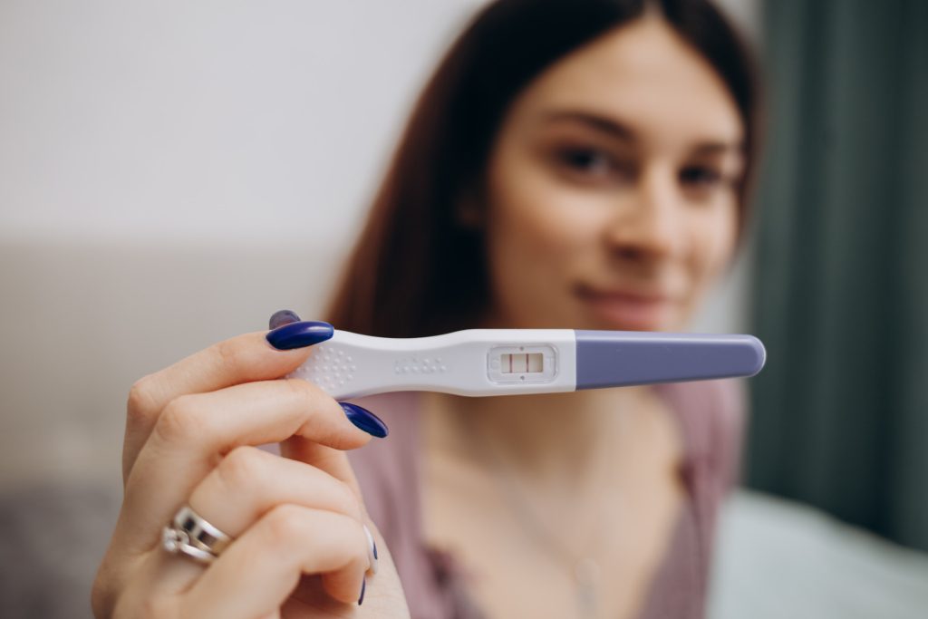
Step 6: Read the test results. A positive result will show two lines, while a negative result will show only one line.

Reading the Results

It is essential to read the results of a pregnancy test within the recommended time, as specified in the instructions. If you wait too long, the test results may not be accurate. Additionally, if you see any faint lines or are unsure of the results, it is recommended to retake the test or confirm the results with a doctor.

Factors That Can Affect the Results of Pregnancy Tests

Medical Conditions
Some medical conditions can affect the results of a pregnancy test. These conditions include ectopic pregnancy, miscarriage, and certain medications like fertility drugs. Hence, it is crucial to consult with a doctor if you suspect that you have any of these conditions, as they can affect the accuracy of the test results.

Timing of the test
If the pregnancy test is taken too early, it may result in a false negative as it takes time for hCG levels to increase to a detectable level. Taking the test 2-5 days before your period is best. You can also wait until a week after a missed period to take a pregnancy test. 

Quality of the test
The accuracy of a pregnancy test can depend on its sensitivity and quality. The sensitivity of pregnancy tests varies; some tests can detect lower levels of hCG in urine compared to others.

User error
The results can be inaccurate if a pregnancy test is not used correctly. Hence, reading and following the instructions carefully and using the test at the right time of day are crucial.

What to do if the Results are Positive

Confirming pregnancy with a Doctor
If the results of a pregnancy test are positive, it is recommended to confirm them with a doctor. A doctor will perform a blood test to confirm the pregnancy and determine the estimated due date. Additionally, a doctor can provide valuable information on prenatal care and what to expect during pregnancy.

First steps after confirming pregnancy
After confirming pregnancy, it is essential to make lifestyle changes to ensure a healthy pregnancy. This includes eating a healthy diet, taking prenatal vitamins, and exercising regularly. Additionally, it is recommended to avoid smoking, alcohol, and certain medications that can harm the baby.

What to do if the Results are Negative

Wait and retest
If a pregnancy test is too early, it may not detect the hormone hCG. So it is best to wait a few more days and then retest.

Follow up with a Doctor
Suppose a pregnancy test is negative, but concerns about a possible pregnancy remain; then it is necessary to follow up with a doctor. They can perform a blood test to check for the presence of hCG, which can detect a pregnancy earlier than a urine test.

Consider other causes
Other reasons besides pregnancy can cause a missed or late period, such as stress, changes in weight, and hormonal imbalances. Therefore, it is essential to consider other possible causes and discuss them with a doctor.

Conclusion

Pregnancy home test kits have made it easier for women to detect pregnancy at home. These tests are easy to use and provide accurate results within minutes. Additionally, it is recommended to confirm the results with a doctor and make necessary lifestyle changes for a healthy pregnancy.

Introduction

Thrombosis comes from the Ancient Greek – thrómbōsis, which means ‘clotting.’ It is the process of the formation of a blood clot inside a blood vessel and is also known as a thrombus. In the process, it causes blocking and obstructs blood flow in the affected area resulting in complications, especially if the clot moves to a critical part of the circulatory system, the brain, or the lungs. 

In the event of an injury of a blood vessel, either a vein or an artery, it is very typical for the human body to prevent further blood loss by using thrombocytes or platelets and fibrin to form a blood clot. When a piece of the blood clot or the clot breaks free, it can form an embolus that moves around the body in the bloodstream, obstructing blood flow to critical organs like the brain or the lungs, resulting in reduced oxygen supply and blood flow with severe complications.

Types of Thrombosis

Now that you know thrombosis occurs when blood clots block blood vessels, you will discover that there are two broad classifications of thrombosis depending on the type of blood vessel affected (arterial or venous) and the exact location of the blood vessel or the organ supplied by it. 

Venous Thrombosis – When thrombosis occurs in the veins or the blood clot blocks a vein, it is called venous thrombosis, also known as DVT, which stands for deep vein thrombosis. In this case, there is a blood clot in the affected part of the body, like the deep veins, axillary or subclavian veins (deep veins of the upper limb), hepatic veins (veins that drain venous blood from the liver), renal vein (the major blood vessel carrying blood from the kidney and the ureter), or jugular veins (veins located within the carotid sheath on either side of the neck). Veins typically from different parts of body to the heart.

Arterial thrombosis – When thrombosis occurs in the arteries, and the blood clot blocks an artery, it is known as arterial thrombosis. Arteries carry oxy-rich blood from the heart to the rest of the body. In the case of arterial thrombosis, the blood supply can be affected and restricted, which can further damage the tissue supplied by that particular artery. 

For example, if arterial thrombosis occurs in a blood vessel in the brain, or what we call arterial embolism, it can lead to a stroke. Similarly, the blood clots could migrate downstream and pose a risk affecting any organ. And if arterial thrombosis occurs in the arteries that supply blood to the heart muscle (coronary arteries), it can lead to a heart attack. The blood clots happen due to the hardening of the arteries or thickening of the artery walls, a condition called atherosclerosis in which there is a buildup of plaque or fatty deposits. This plaque buildup can lead to a rupture followed by a blood clot. Thrombus in the coronary artery can also cause myocardial infarction leading to ischemia. The reduced oxygen supply to the heart cells can often lead to cell death or necrosis. 

Causes of Thrombosis

  • Venous thrombosis can be caused by-

– Immobility

– Any injury or disease to the leg veins

– A fracture

– Being overweight or obese

– Certain medications that can increase the risk of clotting

– Genetic disorders

– Autoimmune disorders.

  • Arterial Thrombosis, on the other hand, is caused by the hardening of the arteries, called arteriosclerosis. This typically occurs when fatty or calcium deposits make the artery walls thick, leading to a fatty buildup of plaque which can burst to cause a rupture, followed by a blood clot.

Generally speaking, some people are predisposed to developing thrombosis and thromboembolism (like newborn babies in the neonatal phase). At the same time, some have a higher risk of developing either venous thrombosis or arterial thrombosis. Also, the chances of development of this condition of thrombosis increase with age and depend on several risk factors and lifestyle factors like the ones discussed above and the following, 

● Heart Condition

● Lung Condition

● Crohn’s Disease (an inflammatory bowel disease affecting the lining of the digestive tract)

● Major Fractures, burns, or injuries to the deep veins in the legs, arms, or the pelvis area

● Lack of movement and physical activity due to travel without moving, medical procedures like surgery or hospitalization, or leg paralysis

● Certain medications like birth control medicines and hormonal contraceptives which contain estrogen or chemotherapy drugs

● Any autoimmune or inflammatory disorder that promotes easy blood coagulation and blood thickening

● Any inherited gene conditions like a family history of blood clots, stroke, or heart attack

● Diet

FAQs:

What is a major cause of thrombosis?

The major cause of thrombosis could be any of the following,

– immobility

– any injury or disease to the leg veins

– a fracture

– being overweight or obese

– certain medications that can increase the risk of clotting

– Genetic disorders

– Autoimmune disorders.

What are the three factors that lead to thrombosis?

Any of the following can be risk factors for thrombosis,

– high blood pressure

– high cholesterol

– diabetes.

What is the most common thrombosis?

The most common thrombosis is arterial thrombosis, which happens in arteries that carry blood from the heart to the rest of the body, often considered to be one of the key reasons for heart attacks and strokes.

What are the signs of thrombosis?

The visible signs and symptoms of thrombosis are pain, swelling, and tenderness in one of the legs, which is usually the calf or thigh, a dull and heavy ache in the affected area, also flushing warm skin in the area of the clot, visibly red skin at the area below the knee and back of the leg.

What is septic arthritis? 

Septic arthritis is a severe infection of joint tissues and fluid caused by germs that may reach the joint from other parts of the body. Such germs travel through the bloodstream. Deep wounds caused at the joints, such as by animal bites, also cause germs to infect them directly. This infection should be treated as soon as it is identified because full recovery is possible. Septic arthritis can be excruciating, progresses rapidly, and can lead to long-term damage to the cartilage and bone of the joint. 

Any joint of the body can be affected by septic arthritis, though more than one is rare. Most commonly, the joints affected by septic arthritis are the knee, hip, shoulder, elbow, wrist, and finger. 

Septic arthritis causes 

You are prone to septic arthritis if your joints get infected by germs and bacteria. Staphylococcus Aureus is the most common bacteria that causes this condition, found even on healthy skin. Besides bacterial infections, septic arthritis can also be caused by viruses or fungi. This condition is also sometimes known as bacterial or infectious arthritis.

This septic arthritis causes of infection can be from:

  • Injury or accident that breaks the skin, like a dog bite, a foreign object going through the skin
  • Complications from a joint surgery
  • Infected wound
  • An open fracture
  • Skin infection or a urinary tract infection spreading to the joints

 You are also at risk of developing septic arthritis if:

  • You have had joint problems like osteoarthritis, rheumatoid arthritis, etc.
  • You have an artificial joint from a knee or hip replacement surgery
  • You have fragile skin caused by issues like eczema and psoriasis
  • You regularly inject yourself with medication causing skin punctures, or inject drugs like heroin.
  • You have a weak immune system caused by HIV, kidney disease, or cancer
  • You have a sexually transmitted disease like gonorrhoea

Other issues that may put you at risk of developing this condition are if you have diabetes, HIV, substance use disorder like alcoholism, are in your late adulthood, or have had septic arthritis before. There are far more severe complications that can be caused by septic arthritis, like an infection of the bone (osteomyelitis), bone death because of poor blood supply (osteonecrosis), limb length discrepancy, sepsis (blood poisoning), chronic pain, and even death. 

Septic arthritis symptoms

Septic arthritis symptoms will manifest differently for different people. Children will express discomfort immediately. They may be irritable or cry if a joint is affected. They may even avoid using the affected joint or halt their usual activity. Besides the symptoms listed below, they may also experience headaches, vomiting, or a sore throat. If parents see a difference in normal behaviour and activity, they should take their child to a doctor.

Commonly found septic arthritis symptoms include:

  • Fever
  • Swelling of the affected joint
  • Pain at the site of infection, which can be severe
  • Loss of appetite and feeling sickly
  • The site of infection is red and hot to touch
  • Inability to move the joint freely or put weight on it

Septic arthritis diagnosis

If you or your child experience any of the symptoms mentioned above, you must seek help from a medical professional. Early diagnosis can prevent much damage from septic arthritis. This condition can lead to blood poisoning if ignored or left untreated. Some essential diagnostic tools can ensure whether the patient has septic arthritis or whether some other serious condition is causing discomfort. They are:

  • Taking down the medical and general health history of the patient.
  • Knowing what the symptoms are and how long they have been present.
  • Draining fluid from the affected joint to analyse what kind of microorganism has caused the infection, the presence of white blood cells, according to which medicines will be given.
  • Blood tests to detect the presence of a bacterial infection.
  • Testing spinal fluid, urine, and phlegm to detect bacteria.
  • Imaging tests like X-rays and MRI to identify how much damage has been caused to the joint and how much it is inflamed.

Septic arthritis treatment

After a diagnosis of septic arthritis is made, there is a possibility of hospitalisation for about two weeks. During this time, you will be given antibiotics through an IV. Antibiotics work fast and can better the symptoms in about 48 hours. For septic arthritis caused by fungi, the doctor will prescribe fungal medication. You will be given medicines for the pain and fever too. The doctor will remove the infected fluid or even pus, if there is any, from the affected joint using a syringe. 

Other ways to do this are with a tube or open surgery if the septic arthritis infection is deep in the joint. You may have multiple fluid removal sessions for the infection to dissipate completely. After being discharged from the hospital, you will be asked to take antibiotics for several weeks. Physical therapy will be recommended to keep your muscles and joints functioning as before the septic arthritis infection. 

You will also be given a splint on the joint to reduce pain. For people who experienced septic arthritis, because their artificial joint got infected, the joint will be removed. The joint will be replaced after the infection goes away.

Final note

We must emphasise that septic arthritis is a major health emergency otherwise; there is a strong chance of irreversible damage. If septic arthritis is detected and treated in time, there will be complete recovery, and there will be little to no lasting after-effects of the condition. When symptoms ease after treatment, you can partake in physiotherapy to keep your muscles, and the joints used to the movement. If you do not do this, you may experience joint stiffness and be unable to use the joint fully.