Category

Surgery:

Category

Introduction

Aortic aneurysm, as the name suggests, happens in the aorta. It is typically linked with the hardening of the arteries, also known as atherosclerosis. This condition may be inherited or a result of complications due to high blood pressure or smoking. Aortic aneurysms start as small bulges which eventually grow in size as the flow of blood puts continuous pressure on the wall of the aorta, the major blood vessel that carries blood from the heart to the rest of the body. They occur when part of a blood vessel’s wall weakens and gives way to the bulge, typically the abdominal aorta. A tear in this weakened area allows blood to escape from it. This is known as an aortic dissection. If the aneurysm occurs in the chest region it is called in Thoracic Aortic Aneurysm (TAA), if it occurs in the abdominal region, it is called Abdominal Aortic Aneurysm (AAA). The most common site in the abdomen is below the area where the aorta gives out branches to supply blood to kidneys, it is called an Infrarenal abdominal aortic aneurysm. But they can also occur in the thoracic region, where there are fewer blood vessels. Aneurysm can be associated with other conditions such as Marfan syndrome, and Ehlers-Danlos Syndrome, hypertension and trauma. It also occurs along with heart valve disease likes bicuspid aortic valve stenosis.  

What Causes Aortic Aneurysm?

A weakening of the aorta or aortic wall causes an aortic aneurysm. The most common causes are atherosclerosis and high blood pressure. This happens due to a combination of factors such as ageing, certain genetic disorders, and certain drugs. This can be present at birth or can develop as a result of disease or injury. Aortic aneurysm is not the same as aortic stenosis, which is when there is constriction of the aorta due to the narrowing of the aortic wall.

Types of Aortic Aneurysms

Aortic aneurysms are classified by their size into small, medium, and large categories. The most severe threat of an aneurysm is that it can burst and cause a stroke or massive bleeding, which in either case is life-threatening. A large aneurysm can affect blood circulation leading to blood clots.

Causes of Aortic Aneurysms

The most common causes are

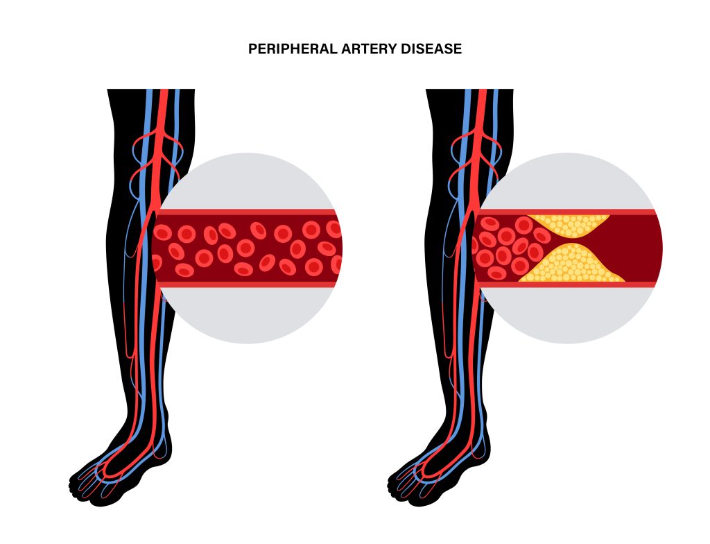
Atherosclerosis- Accumulation of cholesterol in the artery can lead to degeneration of its wall on enlargement. This condition is called atherosclerosis and is the most common cause of aneurysms.

 High Blood Pressure- High Blood pressure puts stress on the wall of aorta. Over the years this stress can lead to bulging of the blood vessel which may be the leading factor for an aneurysm.

Diabetes- Uncontrolled diabetes damages blood vessels by accelerating the process of atherosclerosis, in turn leading to an aneurysm.

Injury / Trauma- An injury to the chest or abdomen can damage the area of the aorta leaving it vulnerable to bulging.

In many cases the cause of aneurysm goes unknown.

Risk Factors involved

Most aneurysms occur in people aged 55 years or older. Although thoracic aneurysms and dissections are known to occur in younger individuals also. Aneurysms are more common in men than women. High blood pressure, smoking and family history.

Symptoms

The symptoms can range from mild to severe, sometimes none at all. But some people are at higher risk of getting one, complete physical examination can help early detection of warning signs.

It’s important to see your doctor right away if you experience any of the following symptoms:

  • Sudden and tearing pain
  • Chest Pain- Often radiating to the back and shoulders.
  • Back Pain – Often mistaken as spine problems.
  • Abdominal Pain
  • Shortness of breath
  • Hoarseness of voice
  • Hemoptysis (coughing up blood)
  • Hemotemesis (Bloody Vomits)
  • Lump near a blood vessel
  • a new bulge in your abdomen, or
  • a sudden change in your heart rate.
  • Aortic regurgitation, when the regurgitation of blood from the aorta causes backflow into the chest. This symptom can be mild or severe.
  • Aneurysm rupture can cause a person’s abdomen to swell causing severe abdominal pain and tenderness which is life threaten and requires immediate emergency care.

Diagnosis

Aneurysm is generally detected by an echocardiogram that can reveal the size and shape of the aorta and if an aneurysm is present. Blood tests can also show an increase in certain proteins that can be present if the aneurysm is large enough to cause blood loss. In a medical diagnosis, your doctor will ask questions on the following,

  • If there is a family history of heart disease
  • Complete physical examination of checking your blood pressure, feel the abdomen for a lump/mass
  • If your doctor suspects an aneurysm, you will be asked to get an ultrasound / echocardiogram / chest x-ray / MR Angiography / CT / Trans esophageal  echo test depending upon the location of aneurysm.

The doctor can also recommend to get a minimally invasive test done known as angiogram.

Treatment of Severe Aortic Aneurysms

Since rupturing of aneurysm is life threatening, it is necessary to get immediate medical attention. Treatment is through medicines or surgery/intervention depending upon site, size and growth rate of aneurysm. Routine testing is recommended to ensure that the aneurysm is not growing. Surgery / Intervention is advised if the aneurysm is growing quickly to avoid a rupture and complications. Aneurysms are unlikely to rupture if they grow slowly and are less than 5.5 cm (about 2 inch) in diameter and causing no symptoms. Serial Ultrasound / CT Scan every 6-12 months to monitor the aneurysm size is the proper treatment for most people along with blood pressure control medicines.

An aneurysm larger than 5 to 5.5 cm (2 inch) requires intervention. Aneurysms that are enlarging rapidly causing symptoms or showing signs of a probable rupture (such as leaking) require immediate medical intervention. Delaying this put an individual at even greater risk of rupture and dead. Newer treatment modalities like endovascular stenting are available to treat large aneurysms using percutaneous techniques avoiding surgery. Stenting is also now providing to be useful in rupture / leaking in advanced centers

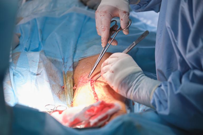
The aneurysm repair aims to strengthen the aortic wall to prevent it from weakening again. An aneurysm repair is usually done under general anesthesia.

Prognosis of Severe Aortic Aneurysms

Generally, people with small aneurysms are not expected to have problems. Most people with aortic aneurysms are expected to have a normal lifespan and not experience any problems with their aorta. However, 10% to 15% of people will have an aneurysm that is large enough to cause significant symptoms, like significant pain or severe swelling. And 1% of people will have a rupture of the aneurysm.

Conclusion

As you now know, an aortic aneurysm is a condition in which part of your aorta – the major blood vessel that carries blood from your heart to your body – becomes enlarged. Call the doctor immediately if you think you may have an aneurysm because if that ruptures, it can be life-threatening. At best, to prevent an aortic aneurysm, you can control your blood pressure, and should you have a family history of heart disease or stroke, then bring changes to improve your health and lifestyle by exercising regularly, watching what you eat, and completely stopping smoking.

FAQS

What are the early symptoms of aortic aneurysm?

Early symptoms of aortic aneurysm include a pulsating feeling in your stomach and nagging back pain.

How serious is an aortic aneurysm?

The aorta is the largest blood vessel in the body which runs from the heart through the center of the chest and abdomen. Therefore, a ruptured abdominal aortic aneurysm can be life-threatening.

Can a person survive an aortic aneurysm?

Some aortic aneurysms are hereditary or congenital, such as bicuspid aortic valve, infection, or inflammatory conditions. You can live with an aortic aneurysm. There are many ways to prevent dissection or a rupture with periodic examinations, medications, and healthy lifestyle.

What happens when you have an aortic aneurysm?

When you have an aortic aneurysm, there will be a bulge or swelling in the aorta, the main blood vessel that runs from the heart down through the chest and tummy. This can get bigger over time and could burst (rupture), can be life-threatening.

What should you not do with an aortic aneurysm?

Your doctor will advise you to avoid heavy lifting and intense physical activities, which can increase blood pressure, putting additional pressure on your aneurysm.

Can you fully recover from an aortic aneurysm?

Typically, you will need 2 to 3 months to fully recover from an aortic aneurysm and be able to resume many of your usual activities after 4 to 6 weeks post treatment.

Suture and Needle choices vary depending on the type of surgery and the patient’s needs. Therefore, knowing the differences between these different types of sutures and when to use them is important to get the best outcome from your surgical procedure. Using the wrong type of suture can lead to undesired surgical outcomes, damage to surrounding tissue or organs, and chronic pain. This can lead to undesired wound healing and complications during recovery, which may require re- suturing for proper wound healing. Suture usage vary on the type of Tissue, patient age as well as the Type of surgical procedure in which suturing needs to be performed. Suturing promotes wound healing by approximating the layers of the tissue and enabling the natural healing process. It also closes the wound thus avoiding the wound to get contaminated by the atmosphere.

What is a suture?

A suture is a surgical device used to close any surgical or traumatic wound requiring skin & tissue closure. A closure is intended to primarily repair skin and deeper layers of the tissue and fascia to fast-track wound healing. Some examples of sutures include monofilament sutures, monofilament polyester sutures, and large absorbable sutures. They are used for repairing cuts and wounds that are deep and open. Sutures are used to close wounds by first suturing the edges together and then using the suture to tie off the wound. After the wound has healed, the suture is either removed or absorbed naturally into the body.

Types of Sutures

Historically, similar to modern sutures, different forms of wound closure mechanisms were employed with needles made of bone and suture materials running from flax, bark, silk, human hair, reeds, etc. Today, there are many options for sutures. Hence, it becomes crucial to make the right suture choice, which means understanding the purpose, attributes, and features of each type of suture. It also means understanding the difference in filament types and the different needle types and under which clinical procedures they have been designed for use. The correct suture helps the surgeon with the right technique and choice of incision to execute and handle the surgical procedure, provide the correct strength, and secure the knots in a manner that can handle and tolerate wound changes during the healing process.

Suture material types are classified as follows:

Absorbable and Non-absorbable Sutures

Absorbable sutures, which get absorbed by the body naturally, are typically employed for deep temporary closure for tissues to heal, and the sutures lose most of their tensile strength over a period of time ranging from weeks to months. Non-absorbable sutures do not get absorbed by the body and hence are required to be removed after a few days of the surgery or may be left in the body permanently. These sutures are used for long-term tissue closures, wounds that take time to heal, like in the case of hernia fascia defects closure. 

Natural and Synthetic Sutures

Sutures are further classified into natural and synthetic sutures. Natural sutures are derived from natural substances. For example Catgut Sutures are usually derived from collagen derived from bovine Guts (used for internal lacerations or wounds). Another Examples of Natural sutures are silk sutures (used in general soft tissue closure and ophthalmic surgery). Catgut degrades by enzymatic reactions (breaking down of proteins partially as peptides or completely as amino acids). They can cause inflammation at the suture site. Synthetic sutures are derived from artificial sources and are made from materials like Nylon sutures (used for general closures and plastic surgery) which is non-absorbable in nature. Synthetic absorbable sutures like Polydioxanone (PDS) sutures (used in abdominal closures or pediatric cardiac procedures) degrade by hydrolysis (the polymer structure of the filaments gets broken down with water penetrating the suture strands) which causes less inflammation at the surgical site. 

Monofilament or Multifilament

Based on the structure, sutures are classified as Monofilament or Multifilament sutures.

Monofilament sutures are single filament sutures with less surface area and higher memory. By memory, we mean the tendency and capacity of a suture to return to its original shape, which makes a suture significantly prone to the knots loosening. They require greater handling and care in the form of more knots to ensure security and less fracture. Monofilament sutures have the capacity to pass through tissues easily, causing less inflammation. 

Whereas Multifilament sutures, as the name suggests, are braided or twisted sutures that are easier to handle by the surgeon as they hold the knots with greater security and less memory. They are prone to inflammation and infection. Multifilament sutures are more expensive than traditional sutures. 

The following are the most commonly used sutures in Surgeries: 

Monofilament sutures, made of a single synthetic strand, are typically used in skin

Closures and Ligation of blood vessels. 

Polypropylene sutures are the synthetic non absorbable sutures; these are typically used in soft tissue closures and are less likely to cause allergic reactions.

Dressings sutures are used to close wounds and stop bleeding. They are typically made of silk, Nylon, or synthetic materials. 

Epithelial sutures are used to close and repair minor wounds. They are made of proteins and are typically used with tissue types such as skin, mucous membranes, and cartilage.

Soft Tissue sutures are used to close wounds in soft tissue such as muscle and tendons. They are also used to support joints and may be made of polyester or polypropylene.

Some sutures are specially used to repair veins and arteries. They are made of polypropylene, Polyester & Nylon.

Needle Types 

The needle is indeed the next most important aspect of sutures after identifying the correct suture type. Needle quality and the appropriate needle choice for a given surgery go a long way to determining the success of the surgery. The surgical needle is mainly built from stainless steel, making it corrosion-resistant. In surgical parlance, the needle has three main parts – the Swage point, the body, and the point. The Swage point is where the surgical suture attaches to the needle by stringing through. The body connects the eye to the point and decides the shape of the needle. In most cases, the needle is curved, but in some cases, it is straight. Curved needles come in different circle lengths like ¼, ½, ⅓, or ⅜ of a circle. The nature of the wound or tissue helps a surgeon choose the needle curve length. The surgeon needs to know where the needle tip is at all times, as it aids in skin closures with minimum trauma. A wrong surgical needle type can lead to tissue injury. The ideal needle is one that is flexible enough to bend yet rigid enough to withstand distortion. It should be sharp to pierce through tough tissues yet thin to cause minimum trauma. Stability of the needle to ensure accurate placement is also equally required. Thusan appropriate surgical needle must be sterile, corrosion-resistant, and firm enough to prevent any bending or distortion, prevent tissue trauma and injury, and ensure easy penetration. 

Different types of needles are based on the tip, like taper needles or cutting ones. Cutting needles have a tip with three sharp edges. In a typical conventional cutting needle, the cutting surface is inside the needle. In contrast, a reverse cutting needle used most commonly for sewing skin has it on the outside. Whereas taper needles are round and can be either blunt or sharp and are recommended for soft and delicate tissues and tendon repair. They typically pierce the skin without cutting it and spread the tissue as it passes through.

Summary

The right type of suture will prevent issues with excessive bleeding, damage to surrounding tissue or organs, and chronic pain. The thickness of the tissue, the location of the tissue, the risk of infection, and the amount of tension, along with different anesthesia requirements and patient age, play an important role in determining which type of suture to use. There needs to be more than the right type of suture to yield the desired surgical results. The correct choice of needle is equally essential and needed for a wound to be sutured with accuracy, precision, and minimal trauma, ensuring fast healing and preventing tissue reaction and inflammation.

FAQs :

Why is it important to use the correct suture technique?

It is important to use the correct suture technique for precision, minimal trauma to the wound at the surgical site and for faster healing.

Why is knowledge of suture material so important?

The knowledge of the right suture material is very important to the surgeon to maximize tissue healing and ensure scar aesthetics and minimal tissue trauma and inflammation at the surgical site.

What factors influence the surgeon’s choice of suture materials?

The main factors that influence a surgeon’s choice of suture materials are

  • Healing time of the wound or tissue
  • Wound type and strength
  • Properties of the suture material
  • Faster attachment of suture and tissue

Common Joint Replacement Myths Busted!

Per the latest statistics published by the Indian Society of Hip and Knee Surgeries Registry, a total of 10,407 hip replacements and 129,371 knee replacements were reported from October 2006 to March 2017 by 150 surgeons.  The demand for joint replacement surgery is on the rise. These surgeries have become more and more common because they offer a quick fix to ailing joints. However, people in line for a  joint replacement surgery often continue to assume a lot about the procedure around pain, medication, recovery and recuperation. Typically, the surgeon will help navigate through the number of questions and doubts the patient may have. Here are some common joint replacement myths that you can quickly fact-check with the help of this article:

Myth – Joint replacement surgery is not permanent.

Fact – In general, many people are afraid of getting knee replacement surgery. They think they have to lie in bed for an extended period after the surgery, which is painful. After surgery, one feels it will hamper day-to-day activities such as walking, running, stair climbing etc. 

These are all misconceptions!

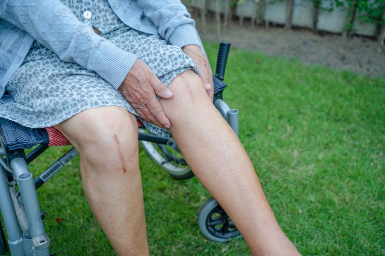
Total knee surgery is a boon for the patient suffering from Osteoarthritis. It improves the quality of life as one gets relief from knee pain and sees a significant improvement in mobility. Knee replacement surgery is one of the most common bone and joint surgeries. 

Knee replacement requires a stay in a hospital. Procedures may vary depending on your condition and your doctor’s practices.

With modern technology, the patient should do activities daily after surgery. One can go walking, can go to washrooms etc. The faster one does it, the better the recovery will be. 

Yes, initially, there will be a little bit of pain, but you can overcome that with time and with the help of a pain management/Physio person.

Myth – You’ll only need physical therapy after joint replacement surgery

Fact – This is another myth that people tend to believe. But, in reality, physical therapy is often recommended for several weeks after joint replacement surgery. For example, knee joint replacement patients are often advised to do static and dynamic stretching exercises for at least two weeks after the surgery is completed. This is because the muscles around the knee need time to heal and get stronger again. Once the muscles have been stretched and strengthened, you can start doing more active exercises like running. Many hip joint replacement patients are also advised to do simple exercises like walking and swimming for a few weeks after the surgery is over. The hip joint is the largest joint in the body. And it’s surrounded by muscles that can be very weak after joint replacement surgery. Therefore, you’ll want to do some simple, gentle exercises to strengthen the muscles around the joints so that they don’t get too weak.

Myth – Joint replacement surgery only works for severe arthritis.

Fact – This is another common joint replacement myth. Most types of arthritis do not lead to severe pain and inflammation that leads to arthritic joints becoming severely damaged. In fact, about one-third of all arthritis cases are caused by other medical conditions or injuries. Joint replacement surgery is performed in these cases as well.

Myth – Joint replacement is only for elderly people.

Fact – This joint replacement surgery myth needs to be busted. Joint replacement surgery is not something that is only recommended for elderly people. In fact, it’s one of the most common surgeries performed today. As a matter of fact, the joint replacement procedure is already available as a third option for treating damaged joints. This means that it can be done in patients of any age.

Myth – Joint replacement is too expensive for person. the average

Fact – This is another myth that’s busted. Joint replacement surgery is one of the safest surgeries that a person can undergo. In fact, some joint replacement surgeries are now offered as a government-funded program for those who meet certain health criteria. These surgeries are typically performed by a doctor who does not charge anything upfront. There are no out-of-pocket expenses like insurance premiums or copayments.

Myth – Joint Replacement Recovery Time Is Incredibly Long

Fact – activities and lead a much more active life. Most people are able to walk just a few hours after joint replacement surgery is completed. They’re not able to run and do more vigorous activities right away. But, within a few days, most patients are back to their normal, active lifestyles. In most cases, full recovery from joint replacement surgery is complete in between one to three months. That’s it! Ready to feel better and not worry about hip or knee pain? Schedule your joint replacement surgery consultation today.

Myth: Bedridden for weeks after the surgery

Fact: This is a false notion. In fact, your surgeon will advise and encourage you to walk or climb a few stairs the same day or the day after surgery. This light activity is suggested to help warm up the muscles and reduce any chance of blot clot formation, which is not good. Typically, hip surgery patients can walk without the walker after a few days and discontinue the use of the walking cane after a few weeks.

Myth: Joint Replacement Surgery means no more sports

Fact: This is not true at all. Initially, during the recovery period, the patient will be asked to stay away from any intensive and strenuous physical sport and activity but they can always pick up other low-impact games and sports like cycling, tennis, golf, swimming and walking.

Overall, most patients report feeling better after joint replacement surgery. They can resume their normal

Hernia surgery is typically a very routine procedure to repair the hernia. In most cases, surgery becomes the only option to treat hernia since it does not get healed on its own. A surgeon performs incisions in the patient’s abdomen, removes the hernia sac or pushes it back into the abdomen, and closes the incision. The process is repeated on another side of the body if needed. The size of the incisions depends on the type of hernia surgery performed. In the case of open surgery, the cut is made at the location of the hernia into the body. In Laparoscopic Surgery, tiny holes are made to insert the surgical tools to perform the surgery. Robotic surgery is a newer technique of hernia surgery. It is similar to Laparoscopic Surgery except that in Robotic Surgery, the surgeon performs the surgical operations with the help of three dimensional images of the abdominal cavity, and robotic arms perform the procedure controlled by the surgeon from his console.

Robotic surgery offers surgeons a new way to approach these operations. There are several advantages of robotic surgery over traditional surgery. The first and foremost advantage is the precision, access to difficult locations inside the cavity, 3D view, motion control, AI-enabled decision making, comfort etc. and many more.

Robotic hernia repair is one of the surgical procedures using a robot to repair a hernia.

Advantages of having new-age techniques Hernia Surgery

Reduced infection: According to several studies, robot-assisted surgery can reduce the risk of infection by up to 80%, which is a common cause of surgical site infection-related death among patients undergoing traditional abdominal surgeries.

Reduced risk of injury: With traditional surgery, surgeons aren’t always sure if they injure a healthy organ. Robotic surgery, on the other hand, doesn’t have these risks.

Fewer surgeries: Another advantage of robotic surgery is the reduction in surgeries performed by surgeons. One study found that robotic hernia repair can cut the need for additional surgeries by as much as 86%.

Greater precision: Robotic Surgery can often achieve greater precision and accuracy than traditional surgery. This can result in less damage to a healthy organ and a shorter hospital stay.

Factors like the cost and availability of robotic systems currently limit their use in many countries. However, these costs may come down in the foreseeable future, allowing surgeons to perform more procedures with robotic technology. Also, robotic systems need to be properly maintained to function properly. This can be a challenge in some countries. The key advantage of robotic surgery is the precision and accuracy provided by the robot. This requires surgeons to use different techniques, which can take some time to master.

Steps followed by surgeons to perform robotic hernia repair

The laparoscope is the key to performing robotic hernia repair. The laparoscope is a thin tube with a camera at the end. A surgeon makes a small abdomen incision and inserts the laparoscope. Once the incision is made, the surgeon can view the abdomen and perform the surgery as if inside the patient’s body. To allow the surgeon to have a good view of the inside of the abdominal cavity, some space is created by inflating the abdomen using harmless gas. The tiny camera attached at the end of the laparoscope projects three-dimensional images of the three dimensional images on the screen. The robotic system used for robotic hernia repair includes a robotic arm and a surgical tool. The surgeon controls the robotic arm using a computer. After the incision is made, the surgeon can control the robotic arm to separate the hernia from the tissues around it. The surgeon gently removes the hernia sac or pushes it back into the abdomen. He then closes the incision.

FAQs

Can a hernia be repaired with robotic Surgery?

Robotic Surgery for Hernia Repair Robotic surgery is a new-age, state-of-the-art technique for repairing hernias where the surgeon is seated at a console and is aided by a robot for precision while the surgeon controls and handles the surgical instruments from the console. The room for error is massively reduced while the process offers three-dimensional images of the abdomen and ensures more minor scars and less pain.

What is the best way to repair a hernia?

With medical technology achieving significant leaps and bounds in terms of innovations with robotic and minimally invasive approaches, surgeons today can perform hernia repair surgery through tiny incisions resulting in quicker healing time, less infection, and more precision, unlike the large incisions associated with traditional Open Surgery.

Wounds can be extremely painful. These wounds can also pose a health risk if they become inflamed or infected. The cause of the suture getting infected could be bacteria, especially those that are naturally found on the skin and Atmosphere. Thus, wound closure may end up promoting bacteria proliferation which can lead to wound complications. As a result, operating doctors, nurses, and other medical professionals have created ways to reduce surgical site infection, speed up healing, and prevent any of these healthcare-associated infections (HCAIs) and surgical site infections (SSIs) from spreading further. One of the ways they have done this is by using antibacterial sutures (ABS) in wound. This closure procedures article explores the advantages of using antibacterial sutures in wound closure procedures. There are various ways and techniques to treat wounds – sutures, staples, non-surgical medications, etc. 

What Is a Suture?

A suture, also known as a stitch, is a very thin, long thread that is twisted together to form a single strand in case of braided structure or comprises of single thread called Monofilament. Suturing is a method of tying off blood vessels to prevent bleeding and hold a wound together till the natural healing process is sufficiently established.

What is an Antibacterial Suture?

Any wound, by and large, are may get contaminated at the time of closure. As a solution to this problem, an antibacterial suture is a surgical innovation which reduces the risk of Surgical Site Infection (SSI). Antibacterial sutures are coated with Antibacterial agents and other agents containing antibacterial properties. They may help in healing wounds faster by significantly reducing the risk of surgical site infections. Why do you need to use an antibacterial suture? We need to use antibacterial suture because bacteria are the leading cause of many types of infections in wounds. You can prevent these infections by covering the wound surfaces with a substance that kills them. An antibacterial suture (Absorbable) is a prosthetic implant into the wound, making it impossible for the bacteria to grow on the surface. You may then cover the wound with a bandage.

Types of Wounds that Can Use Antibacterial Sutures

The first thing you should know about using antibacterial sutures in wound closure procedures is that they can be used on various types of wounds. Antibacterial sutures can be used

  • To close the skin tissue after the surgical procedure
  • To Ligate blood vessels during the planned surgical procedures

In this case, we are referring to regular cuts and injuries & suturing is done in a sterile atmosphere.

How an Antibacterial Suture works ?

Ordinary sutures may pose a risk of infection if required OT Protocols are not followed. Sometimes, an already sutured wound can rupture and burst open if the healing is not complete – this happens due to bacterial infection. If a wound is not sutured correctly, this is an additional complication and cosmetic loss. The Antibacterial coating on the Antibacterial sutures acts as an additional shield against the bacterial growth.

Advantages of using an Antibacterial suture

The layperson needs to know why a surgeon uses an antibacterial or antimicrobial suture technology in their clinical practice. There is enough evidence and data in surgical studies and peer-reviewed journals that an antibacterial suture is a huge clinical benefit. It has a purpose in its design to add value to risk reduction strategies for any SSIs.

  • Prevents Infection – One of the main advantages of using an antibacterial suture is that it prevents infections in a wound. Bacterial infections can be very dangerous. If they go untreated, they can lead to sepsis. Sepsis is an extreme form of blood poisoning that is very dangerous and can quickly lead to death. Preventing these infections means less chance of them spreading throughout your body and causing severe health complications. 
  • Patient Safety – Antibacterial sutures contribute significantly to patient safety by reducing microbicidal activity, which can be both internal and external, enhancing clinical effectiveness and being cost-effective. Antibacterial coated sutures create a Zone of Inhibition around the suture site preventing Bacterial colonization. Hence, antibacterial sutures become the need of the hour to reduce bacterial adherence to surgical sutures.
  • Effective Results – As an invasive innovative technology, antibacterial sutures have stood the test of time ever since they were introduced in the early 1990s. Since their introduction and usage in wound closures, there has been significantly less wound dehiscence, delayed healing, emergence of resistant organisms, toxicity, or allergic reactions.
  • Economically viable – In paediatric and adult surgical procedures, antibacterial suture technology is economically effective and viable. The patient’s Length of Stay may reduce due to the wound being closed with the help of Antibacterial sutures. An added advantage of Antibacterial coating helps in preventing bacterial growth along the line of suture thus preventing unpredicted wound healing.

Conclusion

Globally recognized health authorities like the Centers for Disease Control and Prevention (CDC), the World Health Organization (WHO), and the American College of Surgeons & Surgical Infection Society (ACS & SIS) have recommended the use of antibacterial sutures like triclosan-coated sutures to prevent surgical site infection (SSI). Their guidelines on reducing the risk of SSI are general to antibacterial triclosan-coated sutures, not specific to any one brand. Also, using ABS for skin closure in surgical patients displayed a reduced risk of developing surgical site infections and postoperative complications.