Category

Aortic Stenosis/TAVR/TAVI

Category

The possibility of having heart disease or a heart valve condition is overwhelming and frightening for most people. That is why having an understanding of heart diseases is important so that one can be well equipped with the right information, and hence take the right steps towards treatment.

Heart valve conditions are usually due to birth defects or age-related issues. Many heart valve conditions can be first identified with the help of a murmur, that is a whoosing sound as blood flows from one chamber to the next, or it may sound like an extra click when a valve allows backflow.

A murmur may indicate conditions such as Aortic stenosis, prolapse, regurgitation, etc. While medicines can be prescribed for a heart valve condition, they are mainly for the following reasons

  • To reduce unpleasant symptoms from milder forms of the disorder.
  • To maintain heart rhythm if a related arrhythmia is present.
  • To lower the patient’s risk for clotting and stroke.

Valve diseases are generally progressive by nature, and the outlook for those who do not receive adequate and timely treatment could be fatal. If you are suffering from an aortic valve disease it is highly advisable to opt for a surgical procedure for low surgical risk patients.

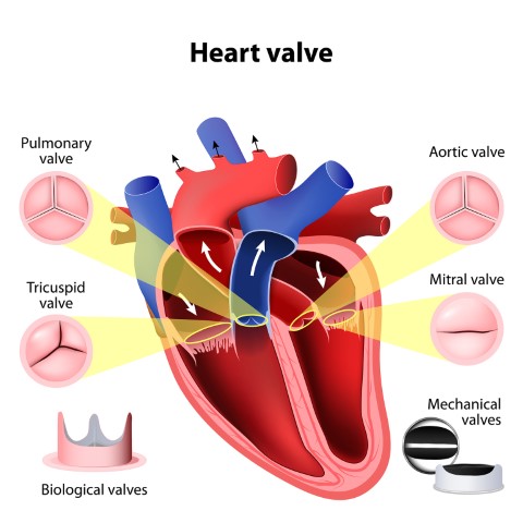
The heart has four chambers. The two upper chambers are called the left and right atrium and the lower chambers are called the left and right ventricle. The four valves that are at the exit of each chamber carry out the function of continuous blood flow through the heart to the lungs and the rest of the body. The four valves are the tricuspid valve, pulmonary valve, mitral valve, and aortic valve.
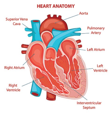
The aortic valve controls blood flow from the left ventricle into the aorta (the main artery in your body). When this valve opens, the oxygen-rich blood is pumped to the aorta and then to the rest of the body. It is key to the body’s blood circulation system.

Reasons you might need to undergo heart valve surgery

Types of aortic valve disease that may require treatment with aortic valve repair or replacement include the following:

Congenital heart disease: This condition may contribute to other complications that prevent the aortic valve from operating properly. For instance, a person may be born with a condition where the aortic valve does not have enough tissue flaps (cusps) or with a valve that is the wrong size or shape, or without an opening that allows blood to flow normally.

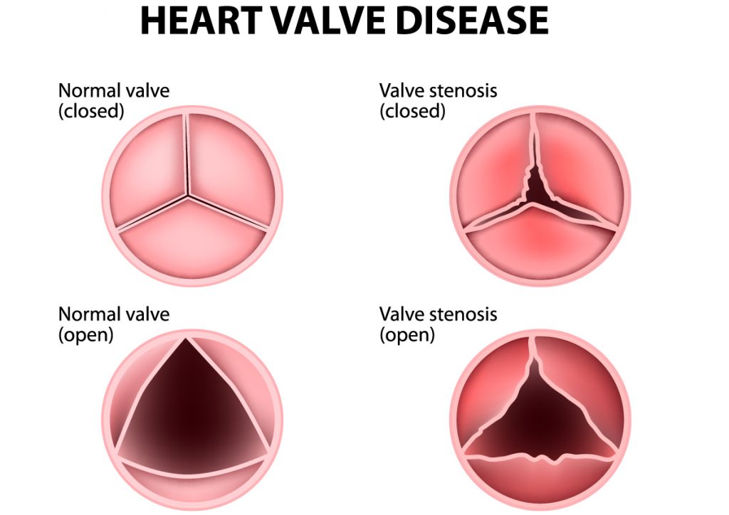
Aortic valve stenosis: When the aortic valve becomes narrow or obstructed, it becomes difficult for the heart to pump blood into the aorta. This may be caused by the thickening of the valve’s closure flaps, congenital heart disease or post-inflammatory changes associated with rheumatic heart disease. Around 75% of patients with unoperated aortic stenosis may die 3 years after the onset of symptoms.

Bicuspid aortic valve: A birth defect where only two cusps grow instead of the normal three. This is a common cause of aortic stenosis. Another cause may be that the valve opening does not grow as the heart does. This makes it harder for the heart to pump blood to the restricted opening. The defective valve becomes narrow and stiff over time because of a calcium build-up. Age-related Aortic Stenosis usually begins after age 60, but often doesn’t show symptoms until ages 70 or 80.

Aortic valve regurgitation: When the blood flows backward through the aortic valve into the left ventricle each time the ventricle relaxes rather than in the normal one-way direction from the ventricle to the aorta. This may be caused due to an abnormal valve shape present at birth (congenital heart disease) or by a bacterial infection.

Most symptomatic patients of AR will require valve replacement surgery within 2 to 3 years of developing breathlessness and symptoms of heart failure. A Swedish study has proven that these individuals have a life expectancy of 2 years lesser than the healthy population of the same age.

Valve diseases, although gradual and progressive are fatal if not treated in the right manner. Valve replacement surgery is recommended as it helps alleviate your symptoms and add quality years to your life.

Careful monitoring and doctor supervision may be all that is needed for a few people with mild aortic valve disease without symptoms. However, in most cases, aortic valve disease and dysfunction can get worse.

Such conditions eventually require surgery to reduce the risk of complications such as heart failure, heart attack, or a stroke or death by cardiac arrest. This is when a doctor will recommend heart surgery.

What are the consequences of not having a heart valve surgery?

  • Decreased life expectancy and quality of life

As per an estimate, as the aortic valve disease gradually worsens, the average rate of survival without undergoing surgery is a mere 50% post 2 years, while it is 20% post 5 years.

While you may feel normal and not notice any problems for years, valve disease is not a condition to be ignored. Once you begin to experience symptoms like the ones mentioned above, not only does life expectancy and quality of life decline but the severity of diseases such as aortic heart stenosis increases.

  • Contributes to more health complications

As the wall of the left ventricle works harder to pump blood through the narrow valve

opening into the aorta, the wall might show muscular thickening, along with the symptoms of aortic stenosis like fatigue and weakness.

The thickened wall allows less room for an adequate amount of blood circulated to the body, as it takes up more space inside the lower heart chamber. This may cause heart failure.

It is advisable to undergo valve replacement surgery as it not only adds to a better quality of life and solves the issue of aortic stenosis but also helps avoid the consequential conditions that come along with it.

Coping with Aortic Stenosis can not only be stressful physically but also mentally. Learning about your condition and the right treatment by talking to your doctor about it can help a great deal. Aortic Stenosis can be fatal and not undergoing the right treatment or prescribed surgery can lead to not just a deterioration in your condition but also create newer complicated conditions for your health. Knowing and understanding your condition as well as the benefits and success rates of the treatment gives you an upper hand and a better chance at survival.

Disclaimer: This blog is only for awareness purposes. We do not intent to promote any medications given in the blog. Please consult your physician before taking any medication.

The human heart has four chambers, with the upper chambers being the left and right atrium and the lower chambers being the left and right ventricles. The heart valves are present at the end of each chamber and they maintain the one-way blood flow from the heart to the lungs and the rest of the body. The four different valves are – Pulmonary Valve, Tricuspid Valve, Aortic Valve and Mitral Valve.

The tricuspid valve is placed between the right atrium and right ventricle. Through this valve, blood flows to the right ventricle. The function of the pulmonary valve is to control the flow of blood between the right ventricle and the lungs. The mitral valve is positioned between the left atrium and left ventricle and allows the blood to flow between them. The aortic valve controls the blood flow from the left ventricle to the aorta. The aorta is the main artery of the body, from which blood flows to the other parts of the body.

What are Valvular Diseases?

Valvular diseases, commonly known as heart valve diseases, occur when one or more of the heart valves do not open or close properly, affecting the blood flow. When more than one valve is affected, it is known as Multiple Valvular Heart Disease. The different types of valvular diseases are:

Stenosis:

In this condition, the heart valves stiffen, leading to the narrowing of the valves. This in turn restricts the blood flow from the valves to the other organs. It keeps the valve from opening fully to allow smooth blood flow, due to which an inadequate amount of blood is supplied to the body. In severe cases, the valve opening may completely close, due to which the blood flow is blocked.

  • Tricuspid valve stenosis: In this condition, the blood is unable to flow from the right atrium to the right ventricle, causing the atrium to enlarge. This affects the blood pressure and flows in the surrounding chambers. It can also cause the right ventricle to shrink or become smaller, restricting the blood circulation to the lungs.
  • Pulmonary valve stenosis: If the pulmonary valve narrows, the blood flow from the right ventricle to the lungs is restricted, due to which oxygen-rich blood cannot be pumped to the rest of the body. In this condition, the right ventricle might have to compensate by pumping blood harder through the narrowed valve, increasing the pressure on the heart.
  • Mitral valve stenosis: In this condition, the blood flow from the left atrium to the left ventricle is reduced, causing fatigue and shortness of breath. It may also cause enlargement of the atrium and fluid build-up in the lungs.
  • Aortic valve stenosis: This is one of the most common heart conditions. In this condition, the blood flow from the heart to the aorta and the rest of the body is restricted. Due to this, the left ventricle has to pump harder to ensure blood flow to the aortic valve. This may also lead to the thickening of the left ventricle.

Prolapse:

Valvular prolapse is a condition in which the valve flaps slip out of place or bulge. This can lead to the valves being unable to close properly. Due to this, blood may flow backwards through the half-closed valve, disrupting the one-way blood flow.

  • Mitral valve prolapse: Also known as Barlow’s syndrome, click-murmur syndrome and floppy-valve syndrome, this condition prevents the valve from closing evenly. The mitral valves may also bulge upwards into the atrium when the two ventricles contracts. Due to this, a small amount of blood may flow backwards through the valve.
  • Tricuspid, Pulmonary and Aortic valve prolapse: These prolapses are less common as compared to Mitral valve prolapse. In the above-mentioned conditions, the leaflets/flaps of the valves do not close properly to form a tight seal.

Regurgitation:

Regurgitation happens when the valve doesn’t close properly, allowing the blood to flow backwards. This disrupts the one-way blood flow and puts a strain on the heart. This reduces the efficiency of the heart in pumping oxygen-rich blood to the rest of the body.

  • Tricuspid valve regurgitation: In this condition, the blood pumped from the right ventricle to the lungs may flow back to the right atrium, leading to the enlargement of the atrium.
  • Pulmonary valve regurgitation: In pulmonary valve regurgitation, the pulmonary valve doesn’t close properly, leading to the blood flowing backwards from the lungs to the heart. This backward blood flow may cause oxygen-rich blood to blend with oxygen-poor blood, reducing the availability of oxygen-rich blood to the rest of the body.
  • Mitral valve regurgitation: Mitral valve regurgitation can be a result of mitral valve prolapse. In this condition, blood leaks backwards into the left atrium through the mitral valve. In severe cases, this condition may lead to the enlargement of the atrium and lung congestion.
  • Aortic valve regurgitation: In this condition, blood leaks backwards from the aorta to the left ventricle, due to which the body does not get an adequate amount of blood, leading to the heart pumping harder to compensate. This may cause the thickening of the left ventricle.

Symptoms of Valvular Heart Diseases:

Some common symptoms of heart valve diseases are:

  • Discomfort or tightness in the chest.
  • Palpitations are a common symptom of heart valve diseases, which may cause the heart to enlarge, leading to irregular heartbeats (arrhythmia).
  • Shortness of breath – as valvular diseases disrupt the flow of oxygen-rich blood to the body, causing breathlessness.
  • Fatigue and weakness
  • Dizziness and fainting
  • Swelling occurs when the blood flows backwards, leading to fluid buildup and congestion, which in turn leads to swollen abdomen and feet.

Though the symptoms may slightly vary for different valvular diseases, it is important to be mindful of them and connect with your doctor even if you experience the mildest of symptoms. Though severe conditions may require surgical procedures, a few lifestyle changes will help keep these heart valve conditions at bay. Be aware and take precautions!

As we move ahead in life, it’s not just our lifestyles that change – the amount of pressure we put on our hearts increases as well. The increased strain at times gives way to various heart diseases, one of the most common ones among them being Aortic Valve Stenosis.

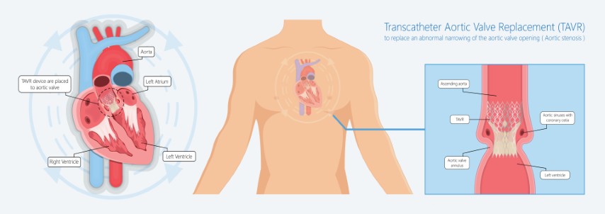
Aortic Valve Stenosis is a condition of the heart where one of the four valves i.e. the aortic valve is narrowed and fails to open properly. This causes various cardiovascular diseases due to inadequate blood supply and pressure on the aortic valve. To treat the said condition, a procedure called Transcatheter Aortic Valve Replacement (TAVR) is performed.

What is a TAVR Procedure?

TAVR is a minimally invasive medical procedure that is used to treat Aortic Stenosis (AS). Before TAVR, Surgical Aortic Valve Replacement (SAVR) or open-heart surgery was the only way to cure aortic stenosis, but the procedure also led to several other complications among patients. But TAVR has made this procedure Less painful, less scarring with a faster recovery.

As this approach avoids surgically opening the chest or making an incision on the heart, TAVR is a  safer option for high-risk patients. Before deciding on undergoing TAVR, you must consult your cardiac surgeon and specialists to determine if the procedure is beneficial for you. 

Why does Aortic Valve Stenosis need to be treated?

A few of the complications that arise with Aortic Stenosis are chest pain, extreme fatigue, fainting, gasping, and swelling on the leg. It may also lead to cardiac arrest and heart failure. Hence, it must be treated immediately.

What has involved in the Transcatheter Aortic Valve Replacement (TAVR) procedure?

A catheter, which is a small, flexible tube is used to place a new valve in the heart. The doctor will make a small incision in a blood vessel in your leg or the chest wall. Once done, the doctor uses advanced imaging techniques to guide the catheter to your heart and into the aortic valve and position it properly.

What are the different types of TAVR Procedures?

While TAVR is essentially the placing of a valve via a catheter, the procedure varies from case to case. These include –

Transfemoral TAVR: A small opening is made to access an artery in the groin area at the top of the leg.

Transapical TAVR: An incision is made in the chest wall, between the left ribs, through which the catheter is inserted.

Transaortic TAVR: A small opening is made on top of the breast bone, through which the catheter is directly inserted into the aorta. 

Transcaval TAVR: An incision is made in a large vein in the groin area, through which the catheter can be inserted, from which it goes through the vena cava to the abdominal aorta, and into the aortic valve. 

Why can’t all Patients with Aortic Valve Disease opt for a Transcatheter Aortic Valve Replacement (TAVR) procedure?

One can opt for TAVR if they have Aortic Stenosis that leads to other symptoms and complications. They can usually opt for TAVR if they are at a higher risk of experiencing complications from open-heart surgery. Open-heart surgery is also risky if one has a kidney or lung disease. Though TAVR might be preferred due to its minimally invasive nature, one must be thoroughly evaluated before undergoing a TAVR. Patients with extremely high risk to moderate risk conditions are usually the ideal candidates for TAVR. Similarly, patients with active endocarditis infection or a similar aneurysm also cannot opt for a TAVR. Bicuspid patients who don’t prefer an open-heart surgery can seek a thorough evaluation with the TAVR team to discuss the risk factors and benefits and to also seek other treatment options. Also, patients in their 50s and 60s are refrained from opting for TAVR due to limited data present for the durability of bioprosthetic heart valves.

Are there any risks involved in a TAVR procedure?

Every medical procedure comes with some risks and the same applies to TAVR too. Hence it is important to have an expert opinion before undergoing any surgery for Aortic Valve diseases. Some of the major risks involved in TAVR are as follows:

Bleeding: Though the procedure is minimally invasive, it involves inserting a catheter in your leg, which may lead to internal bleeding.

Complications in the blood vessels: Injuries are caused due to vascular access which in turn, can lead to tissue malperfusion. Passing catheters through the arteries can also damage them. 

Replacement valve: It might also lead to problems with the replacement valve such as blood leaks around the new valve or the valve slipping out of place.

Arrhythmia: It might lead to irregular heart rhythms, which would require pacemaker implantation.

Stroke: A small percentage of patients undergoing TAVR have complained of stroke, either during the procedure or after it. 

Damage to the kidney: The dye that is used for imaging can affect the kidneys, leading to kidney damage. But this complication is usually reversible.

Heart Attack: A minimal percentage of patients have also had heart attacks during the procedure.

Infection: Passing catheters through the arteries can also lead to infections, but they are usually treatable.

Death: While TAVR is usually effective, there is a very low possibility that the patient cannot survive the procedure. 

When it comes to heart health, it is important to weigh the pros and cons properly to avoid health issues in the future. As technology advances, the risks associated with it grows. But proper medical care and procedure surely avert all the risks and helps people to lead a normal life after TAVR or any surgery. All you need to do is take proper precautions and follow the advice of your doctor in the pre and post phases of the surgery to keep your heart healthy in the long run.

Apart from being an extremely vital organ, the heart is also a complicated one! Over the years, we have learned a lot about matters of the heart and many ailments can now be treated medically. One such ailment is Aortic Stenosis.

So, what is Degenerative Aortic Valve Disease?

Degenerative Aortic Valve Disease, also known as Degenerative Aortic Stenosis, is usually found among patients who are above 60 years of age. It is one of the most common cardiac diseases after hypertension and coronary heart disease.

Degenerative Aortic Valve Disease, also known as DAVD, is a condition in which the left ventricle (the main pumping chamber in the heart) and the aorta don’t work properly, leading to complications. It is most prevalent in older people with a tricuspid valve, though younger people with a bicuspid valve are also susceptible to it. There are two types of Aortic Valve Diseases:

Aortic Valve Stenosis: In this condition, the cusps (leaflets) of the aortic valve become thick and stiff, which leads to the narrowing of the valve opening. This in turn blocks or restricts the flow of blood from the heart to the rest of your body.

Aortic Valve Regurgitation: In this condition, the valve doesn’t close properly, causing the blood to flow back to the left ventricle, which may prevent the heart from efficiently pumping blood to the rest of your body.

The Symptoms of Aortic Valve Disease may be mild or may not be experienced for many years. Some of the most common symptoms are:

  • Abnormal heart sound
  • Gasping and shortness of breath
  • Fainting
  • Tightness in the chest
  • Extreme fatigue
  • Arrhythmia (Irregular heartbeat)

What causes Degenerative Aortic Valve Disease?

The heart has four valves that aid in the blood flow. Each valve has a flap that opens or closes during every heartbeat. In Degenerative Aortic Valve Disease, the valve between the left ventricle and the main artery doesn’t work properly. The valve may either not open fully, blocking the blood flow from the heart to the body (stenosis), or the valve may not close properly, causing the blood to flow backward (regurgitation).

Aortic valve disease may be caused due to a heart defect present from birth, or due to other factors such as age, high blood pressure, infection/injury to the heart.

Risk Factors of Degenerative Aortic Valve Disease

Old Age: As mentioned before, Degenerative Aortic Valve Disease is more prevalent in older people as compared to young people.

Congenital Heart disease: If you had certain heart conditions at birth, the chances of getting Degenerative Aortic Valve Disease increases.

Infection: If you have had a history of infections in the heart, it may act as an impending factor.

Radiation Therapy: If you have undergone radiation therapy to the chest area, you are prone to Aortic Valve Disease.

Endocarditis: It is an inflammation in the heart’s inner lining involving heart valves, which in turn can turn into a major risk factor.  

What are the complications involved in Degenerative Aortic Valve Disease?

While we now know about the various risk factors, we need to be mindful of the various ways the ailment can affect us. These include –

  • Heart Failure: Aortic Valve Disease may lead to heart attack or cardiac arrest, which in turn will lead to heart failure
  • Stroke: Patients who have Aortic Valve Disease are susceptible to stroke
  • Blood clots: When diagnosed with Degenerative Heart Disease, there is a high risk of blood clots in the heart valves.
  • Arrhythmia: Abnormal heartbeat is a very common symptom of DAVD
  • Death: If not treated properly, on time, Aortic Valve Disease may cause malfunctioning of the heart, leading to certain death.

Can Degenerative Aortic Valve Disease be diagnosed?

After discussing the signs and symptoms with your doctor, he/she may review your medical history and even conduct a physical examination. After the physical examination, your doctor may prescribe certain tests to diagnose your condition.

Echocardiogram: This test uses sound waves to provide images of your heart to evaluate the chambers, aortic valve, and blood flow.

Electrocardiogram (ECG): Electrodes are attached to your skin to measure your heart activity. An ECG can deduct enlarged heart chambers, abnormal rhythms, and other conditions.

X-ray: A chest x-ray can help determine if the heart is enlarged and it also helps doctors examine your lung condition.

Cardiac CT scan: A CT scan uses multiple x-rays to create a detailed image of your heart and valves.

Can Degenerative Aortic Valve Disease Be Treated?

The treatment for degenerative aortic valve disease depends on the severity of your condition. Once you have been thoroughly evaluated, your doctor will discuss with you to provide the right treatment. Some common treatment options are:

Aortic Valve Repair: To repair the aortic valve, surgery needs to be conducted to either separate valve cusps that have fused, removing excess valve tissues, or patching holes in the valves. If diagnosed with Aortic Stenosis, the doctor might conduct a procedure to insert a catheter with a balloon into an artery via the groin and move it towards the aortic valve. The balloon inserted is inflated to expand the valve. Once the valve is expanded, the balloon is deflated, and the catheter is removed.

Aortic Valve Replacement: This is one of the most sought-after measures to treat DAVD. In Aortic Valve Replacement, the surgeon removes the damaged valve and replaces it with a new valve. The new valve can either be a mechanical valve, or a valve made from cow, pig, or human heart tissue. This is usually done with open-heart surgery also known as Surgical Aortic Valve Replacement.

Transcatheter Aortic Valve Replacement: TAVR is a minimally invasive method to treat a narrowed aortic valve that fails to open properly. It is performed on patients after thorough consultation from their doctors. In this procedure, the doctor inserts a catheter through the blood vessel in the leg or chest lining and guides it to the heart to relieve the problem of aortic valve stenosis.

Degenerative Aortic Valve Disease is among the common cardiac diseases, which, if left untreated, can become fatal. While seeking out treatment options, consult with your doctor about which treatment procedure would work best for you. It is also important to weigh the pros and cons of each option before going ahead with the treatment. You can also talk to your doctor about the lifestyle changes you can make to overcome the complications and ensure that the condition doesn’t relapse.

As we all know that our heart is the center of the circulatory system. It is a network of vessels that delivers blood to every part of the body. To tell you more about how our heart functions, let us know more about the four valves that make the whole functioning possible. The heart has four valves – mitral valve, tricuspid valve, pulmonic valve, and aortic valve. 

The mitral valve and tricuspid valve are located between the upper heart chambers and lower heart chambers and amidst the ventricles and the major blood vessels, located are the aortic valve and pulmonic valve.

FUNCTIONS OF THE VALVES

These valves have different roles that keep the heart functioning properly. Hence, it should be healthy and functioning. It must be open all the so that blood can pass and valves must close tightly so no blood flows into another chamber. 

When we talk about the aortic valve, it separates the left ventricle from the aorta, allows blood to leave the heart from the left ventricle through the aorta and the body, and prevents the backflow of blood from the aorta to the left ventricle. 

When the aortic valve isn’t working properly, it becomes difficult for the heart to send blood to the rest of the body which leads to various aortic valve diseases.

AORTIC VALVE DISEASE

There are two major types of aortic valve diseases:

Aortic Stenosis

Aortic Stenosis is the narrowing of the Aortic valve opening. The narrowing of the valve could lead to reduction or blockage of the blood flow from the heart into the main artery to the body and towards the rest of the body. This forces the heart to work harder.

Aortic Regurgitation

Aortic regurgitation or insufficiency causes when the valve doesn’t close properly causing the blood to leak into the heart which over time makes the heart stretch and enlarge which later can lead to heart failure.

AORTIC VALVE TREATMENT

Treating the dysfunction of the aortic valve depends upon the severity of the aortic disease. Traditionally, Aortic Valve Replacement Surgery was done through traditional open-heart surgery. This procedure involves a cut in the chest which is risky and requires a longer recovery time. Another way is replacing a valve via a minimally invasive method where an incision is made in the chest or a catheter inserted in the leg or chest. It is by far the safest option and this procedure is called TAVR or Transcatheter Aortic Valve Replacement. 

This can be done using a mechanical valve or a biological valve. 

WHAT IS A BIOLOGICAL VALVE?

While performing the TAVR procedure, the doctor removes the damaged valve and replaces it with a mechanical valve or biological valve. Biological valves are also known as bioprosthetic valves. That is a valve that is made an animal tissue or human heart tissue. Animal tissue is known as Xenograft and human heart tissue is known as Homograft. Animal tissues are made from a cow or pig. But, to perform this procedure, depends on the severity of the condition and the situation of the patient. Accordingly, if needed, the doctor might suggest replacing the damaged aortic valve with a biological valve. 

The reason why the biological valve is used is that they degenerate over some time and they comparatively require no blood-thinning medicines, post-surgical care, and hospital stay.

Biological valves are not as strong as mechanical valves and they need to be replaced every 10 years or so. These valves breakdown much faster in children and young adults, so these valves are used mostly in elders.

WHAT IS A MECHANICAL VALVE?

Titanium and carbon are the materials used to make Mechanical valves. These valves are sewn in the place of original valves, they consist of two leaflets and a metal ring surrounded by a ring of knitted fabric. The advantage of these valves is that they are durable and last longer. But the valves provide a surface on which it is easy to form a blood clot.

These valves should be avoided for women of child-bearing age and those with a high risk of falls or bleeding.

HOW IS TAVR PROCEDURE PERFORMED USING A BIOLOGICAL VALVE?

TAVR procedure is a minimally invasive procedure that is used to repair or replace the damaged aortic valve. On the day of surgery, the doctor will decide which type of anesthesia is best for you but your heart will continue to beat during the procedure. Once you are asleep, a short hollow tube called a sheath is inserted into your femoral artery. The new valve is placed in the delivery system using a balloon at the end of the catheter. It contains the replacement valve folded around it. This delivery system is pushed up to your aortic valve. 

Once it reaches the diseased valve, it is inflated which pushes aside the leaflets of your diseased valve. This valve replacement surgery usually does not require cardiopulmonary bypass. Once the biological valve is placed, the balloon is deflated and the catheter is removed. 

The doctor will check if the new valve is opening and closing like the normal aortic valve should and close the incision once the new biological valve starts functioning properly. 

PROS OF USING A BIOLOGICAL VALVE DURING TAVR

The benefits of replacing the damaged Aortic Valve with A Biological Valve is that they don’t come with a higher risk of a blood clot. They are the best for people over the age of 65, pregnant women, and people who have kidney stones.

Valves used from Bovine (cow) is not an actual valve but it is a tissue surrounding the cow’s heart. It is strong and flexible. 

Using a biological valve during TAVR operation for pregnant women is more beneficial because it doesn’t require using blood thinners as consuming those medicines can harm the fetus. 

CONS OF USING A BIOLOGICAL VALVE DURING TAVR

TAVR, of course, includes some risks. Generally, risks are minimal which includes complications in blood vessels, infection – if not taken care of it properly, etc. Only in extreme cases, does one suffer from stroke if post-surgical care is not taken properly. It all depends on one’s health and medical history. So, if you or somebody you know is suffering from post-surgical complications, it is better to consult a doctor immediately.

To conclude, it is important to understand the risk and advantages of valve replacement surgeries. It is better to listen to the advice of your doctor and research about TAVR in depth before undergoing the procedure. TAVR is a procedure of the heart, all the precautions must be taken into consideration. And after the TAVR procedure, you should constantly be in touch with your doctor for regular health check-ups.